Sheep Kidney Anatomy: The Ultimate Visual Guide
Understanding sheep kidney anatomy is crucial for veterinary students and professionals alike. Histology, the study of tissue structure, provides a detailed view of the organ’s cellular composition. The glomerulus, a key component within the kidney, functions as the primary filtration unit. Institutions such as the University of Veterinary Medicine often utilize sheep kidney anatomy models for educational purposes, demonstrating the intricate structure of this vital organ and contributing significantly to the understanding of renal physiology.
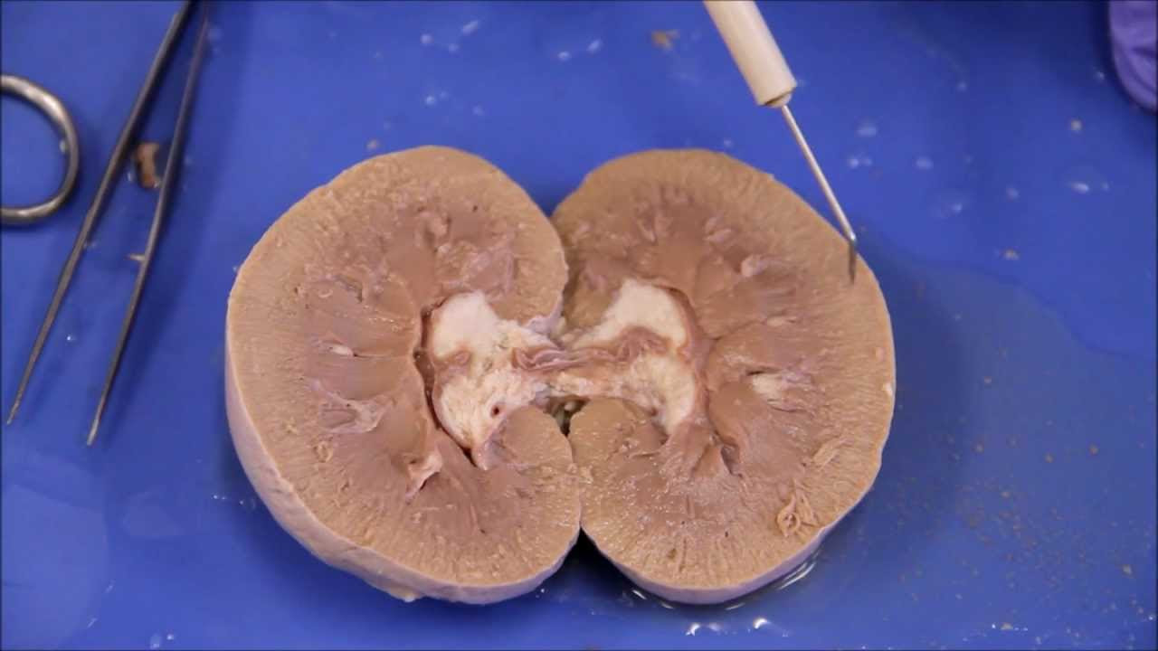
Image taken from the YouTube channel NAUBIO202 , from the video titled NAU Bio 202 Lab 7 – Sheep Kidney Dissection .
Crafting the Ultimate Visual Guide to Sheep Kidney Anatomy
This guide outlines the optimal structure for an article focused on "sheep kidney anatomy," aiming to be both informative and visually engaging. The article will leverage the main keyword, "sheep kidney anatomy," throughout the content to optimize for search engines and ensure clarity for the reader.
Introduction: Setting the Stage
-
Headline: Compelling and keyword-rich (e.g., "Unveiling Sheep Kidney Anatomy: A Detailed Visual Guide"). Consider variations like "Sheep Kidney Anatomy Explained: The Ultimate Illustrated Breakdown."
-
Introductory Paragraph: Briefly introduce the sheep kidney and its relevance. Why should the reader care about sheep kidney anatomy? (e.g., veterinary studies, comparative anatomy, understanding mammalian kidneys). Specifically mention the target audience (e.g., students, researchers, veterinary professionals). Directly address the main keyword "sheep kidney anatomy."
-
Purpose Statement: Clearly state the article’s objective – to provide a comprehensive and visually rich understanding of sheep kidney anatomy. Mention the types of visuals included (diagrams, photographs, potentially 3D models).
External Morphology: The Kidney’s Surface
-
Heading: Sheep Kidney Anatomy: External Morphology
-
General Shape and Size: Describe the typical shape (bean-shaped) and size of a sheep kidney, possibly providing average dimensions. Include a high-quality photograph or illustration of the external kidney. Label key features:
- Renal Hilum: Mention its location and what enters/exits through it.
- Renal Capsule: Describe its appearance and function (protection).
-
Orientation: Explain how to identify the cranial (anterior) and caudal (posterior) ends.
-
Surface Features: Note any observable surface features and their significance.
Internal Structure: A Journey Inside
-
Heading: Sheep Kidney Anatomy: Internal Structure
-
Cross-Sectional Anatomy: The core of this section relies heavily on visuals. Include a well-labeled diagram and a photograph of a bisected sheep kidney. Use consistent color-coding throughout all visual aids for each structure.
Major Regions: A Layered Approach
- Renal Cortex:
- Description: Outer region, granular appearance.
- Function: Site of filtration.
- Visual Emphasis: Clearly delineate the cortex in all diagrams and photographs.
- Renal Medulla:
- Description: Inner region, striated appearance.
- Function: Concentration of urine.
- Visual Emphasis: Show the renal pyramids and columns.
- Renal Pyramids:
- Description: Cone-shaped structures within the medulla.
- Function: Contain collecting ducts.
- Visual Emphasis: Clearly label and differentiate between pyramids.
- Renal Columns:
- Description: Cortical tissue extending between pyramids.
- Function: Provides a route for blood vessels.
- Visual Emphasis: Differentiate the columns from the pyramids and cortex.
- Renal Pyramids:
- Renal Sinus:
- Description: Space containing the renal pelvis, calyces, and blood vessels.
- Function: Collects urine and houses vessels.
- Renal Pelvis:
- Description: Funnel-shaped structure that collects urine.
- Function: Channels urine to the ureter.
- Visual Emphasis: Show how the calyces drain into the renal pelvis.
- Major and Minor Calyces:
- Description: Cup-shaped structures that collect urine from the renal papillae.
- Function: First point of urine collection.
- Visual Emphasis: Clearly distinguish between major and minor calyces.
- Major and Minor Calyces:
- Renal Cortex:
Vasculature: Blood Supply to the Kidney
-
Heading: Sheep Kidney Anatomy: Vasculature
-
Diagram of Renal Blood Flow: Create a detailed diagram illustrating the flow of blood through the kidney.
-
Arterial Supply:
- Renal Artery: Origin and entry point into the kidney.
- Segmental Arteries: Branches of the renal artery.
- Interlobar Arteries: Travel between the renal pyramids.
- Arcuate Arteries: Branch along the base of the renal pyramids.
- Interlobular Arteries: Extend into the cortex.
- Afferent Arterioles: Lead to the glomeruli.
- Glomeruli: Capillary network within Bowman’s capsule (part of the nephron).
- Efferent Arterioles: Carry blood away from the glomeruli.
- Peritubular Capillaries: Surround the tubules in the cortex.
- Vasa Recta: Straight capillaries that follow the loops of Henle in the medulla.
-
Venous Drainage:
- Interlobular Veins: Drain the peritubular capillaries.
- Arcuate Veins: Receive blood from interlobular veins.
- Interlobar Veins: Travel between the renal pyramids.
- Renal Vein: Drains blood from the kidney.
The Nephron: Functional Unit of the Kidney
-
Heading: Sheep Kidney Anatomy: The Nephron
-
Introduction to the Nephron: Explain that the nephron is the functional unit of the kidney.
-
Diagram of a Nephron: Provide a detailed, labeled diagram of a nephron, highlighting its key components.
Nephron Components: A Detailed Breakdown
- Renal Corpuscle:
- Glomerulus: Capillary network where filtration occurs.
- Bowman’s Capsule: Surrounds the glomerulus and collects filtrate.
- Renal Tubule:
- Proximal Convoluted Tubule (PCT): Reabsorption of water, glucose, amino acids, and ions.
- Loop of Henle: Concentrates the urine.
- Descending Limb: Permeable to water.
- Ascending Limb: Impermeable to water, actively transports ions.
- Distal Convoluted Tubule (DCT): Further reabsorption of water and ions, secretion of waste.
- Collecting Duct: Collects urine from multiple nephrons.
- Renal Corpuscle:
-
Juxtaglomerular Apparatus (JGA): Briefly describe its location and function in regulating blood pressure and filtration rate.
Sheep Kidney Anatomy: Frequently Asked Questions
Here are some common questions regarding sheep kidney anatomy and the information presented in our guide.
What is the hilum of the sheep kidney?
The hilum is the indented area on the medial side of the sheep kidney. This is where the renal artery, renal vein, and ureter enter and exit the kidney. Understanding the hilum’s location is key to understanding sheep kidney anatomy.
What are the major regions of the sheep kidney?
The sheep kidney is composed of three major regions: the renal cortex (outer layer), the renal medulla (inner layer), and the renal pelvis (funnel-shaped collecting area). These regions perform specific functions in urine production.
How does a sheep kidney differ from a human kidney?
While the basic functions are similar, sheep kidneys are multilobar, meaning they are composed of multiple distinct lobes. Human kidneys, on the other hand, typically have a smoother, bean-shaped appearance without clear lobes. This difference is a key feature of sheep kidney anatomy.
What is the function of the ureter in the sheep kidney?
The ureter is a tube that carries urine from the renal pelvis of the sheep kidney to the urinary bladder. It’s an essential part of the excretory system, ensuring waste products are removed efficiently.
Alright, that’s a wrap on sheep kidney anatomy! Hope you found it helpful. Now go forth and impress someone with your newfound knowledge!
