Noncapsulated Lipoma: What Makes it Different? | Experts
The diagnostic journey for soft tissue masses often leads clinicians to consider various benign neoplasms, where lipomas represent a common entity. While conventional lipomas typically exhibit a well-defined capsule, the nuanced presentation of noncapsulated capsulated lipoma presents unique diagnostic and management challenges. This is where advanced imaging techniques, such as Magnetic Resonance Imaging (MRI), become invaluable, offering detailed visualization of tissue characteristics and anatomical relationships. Expert consultation with pathologists specializing in soft tissue tumors is crucial for accurate differentiation, as they possess the expertise to interpret histological findings and guide appropriate clinical decision-making. Understanding the distinguishing features of a noncapsulated capsulated lipoma is critical for avoiding misdiagnosis and ensuring optimal patient care.
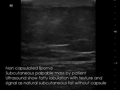
Image taken from the YouTube channel Educational Radiology Channel ERC , from the video titled CASE 661 Non capsulated lipoma .
Noncapsulated Lipoma: Differentiating It from Encapsulated Counterparts
This article aims to clarify the characteristics that distinguish a noncapsulated lipoma from its more common, encapsulated counterpart. Understanding these differences is crucial for accurate diagnosis and appropriate treatment planning. While the main keyword is "noncapsulated lipoma," we will also address the crucial concept of an "encapsulated lipoma" for effective comparison.
Defining Lipomas: A Quick Overview
Lipomas are benign (non-cancerous) tumors composed of fat cells. They are generally slow-growing and often located just beneath the skin, making them palpable as soft, movable lumps. While generally harmless, they can sometimes cause discomfort or cosmetic concerns, leading individuals to seek medical attention. The primary differentiator between types of lipomas lies in whether or not they possess a capsule.
The Importance of the Capsule
The presence or absence of a capsule is a key diagnostic feature. A capsule is a well-defined outer layer of connective tissue that surrounds the fatty tissue of the lipoma. Think of it like the skin of a sausage, neatly containing the contents within.
Encapsulated Lipomas: A Well-Defined Boundary
- Characteristics:
- Clearly defined borders.
- Easily separable from surrounding tissues during surgical removal.
- Lower risk of recurrence after complete excision due to the defined margins.
- How it Works: The capsule acts as a barrier, preventing the lipoma from infiltrating into adjacent tissues. This makes diagnosis relatively straightforward using imaging techniques such as ultrasound or MRI.
Noncapsulated Lipomas: Lacking a Defined Edge
- Characteristics:
- Lack a distinct outer layer or capsule.
- Merge gradually with surrounding tissues (muscle, fat).
- More difficult to surgically remove completely.
- Higher risk of recurrence compared to encapsulated lipomas.
Identifying Noncapsulated Lipomas: Diagnostic Challenges
Diagnosing a noncapsulated lipoma can be more challenging than identifying an encapsulated one because of the lack of clear boundaries.
- Palpation: While both types may feel like soft lumps, a noncapsulated lipoma might feel less defined and more diffuse.
- Imaging (MRI & CT Scans): These imaging techniques are crucial for evaluating lipomas. However, identifying noncapsulated types requires careful interpretation, as the margins may be indistinct, blurring into the surrounding tissues.
- Biopsy: A biopsy, involving the removal of a small tissue sample for microscopic examination, is often necessary to confirm the diagnosis and rule out other conditions. The pathology report will specifically mention the presence or absence of a capsule and the pattern of fat cell distribution.
Treatment Options and Considerations
Treatment for both encapsulated and noncapsulated lipomas usually involves surgical removal. However, the surgical approach and the likelihood of success differ significantly.
- Encapsulated Lipomas: Surgical excision is typically straightforward. The surgeon can easily identify and remove the entire lipoma, including the capsule, with minimal disruption to surrounding tissues. Recurrence is relatively uncommon.
- Noncapsulated Lipomas: Due to the lack of a capsule and the infiltrative nature of these lipomas, complete surgical removal can be challenging. Surgeons must carefully dissect and remove as much of the lipoma tissue as possible without damaging surrounding structures. Because it’s harder to distinguish the lipoma from the healthy tissue, a wider excision might be necessary. This can lead to a higher risk of complications or a greater cosmetic impact. In some cases, complete removal is not possible, increasing the likelihood of recurrence.
Recurrence Rates: Why Noncapsulated Lipomas Pose a Higher Risk
The primary reason for the increased recurrence rate of noncapsulated lipomas is the difficulty in achieving complete surgical excision. Microscopic extensions of the lipoma tissue may remain behind, leading to regrowth over time. Regular follow-up appointments with your doctor are essential after surgery to monitor for any signs of recurrence.
The following table summarizes the key differences between encapsulated and noncapsulated lipomas:
| Feature | Encapsulated Lipoma | Noncapsulated Lipoma |
|---|---|---|
| Capsule | Present | Absent |
| Margins | Well-defined | Ill-defined, blending into surrounding tissues |
| Surgical Removal | Typically straightforward; complete excision possible | More challenging; complete excision may not be possible |
| Recurrence Risk | Low | Higher |
| Diagnostic Challenge | Generally easier | More challenging |
FAQs: Noncapsulated Lipomas
This FAQ section aims to clarify some common questions about noncapsulated lipomas and how they differ from regular lipomas. We hope these answers provide useful information about this specific type of growth.
What exactly is a noncapsulated lipoma?
A noncapsulated lipoma, unlike a typical lipoma, lacks a distinct, fibrous capsule surrounding the fatty tissue. This means the fatty tissue blends more diffusely with the surrounding tissues, making it potentially more difficult to define and remove surgically.
How does a noncapsulated lipoma differ from a capsulated lipoma in terms of appearance and detection?
Capsulated lipomas are typically well-defined and feel like a distinct lump under the skin. A noncapsulated lipoma, however, might feel less defined and have less distinct borders due to the absence of a capsule. This can sometimes make it harder to detect through physical examination alone.
Why are noncapsulated lipomas often more challenging to remove?
Because noncapsulated lipomas lack a well-defined border, it’s more difficult to distinguish them from the surrounding tissue during surgery. This can increase the risk of incomplete removal and potential recurrence compared to removing a regular, capsulated lipoma.
What are the potential complications associated with noncapsulated lipoma removal?
Potential complications following noncapsulated lipoma removal include a higher chance of recurrence if the entire growth isn’t removed. Other risks, similar to any surgical procedure, can include infection, bleeding, and scarring. It’s important to discuss these potential complications with your surgeon.
Hopefully, this deep dive into what makes a noncapsulated capsulated lipoma different has shed some light on the subject. If you’re curious about any of this, or just want to chat more about lipomas, don’t hesitate to reach out! Always remember to consult your doctor for any health concerns.
