Non-Encapsulated Lipoma Removal: Is It Possible?
The treatment of infiltrative lipomas, characterized by their diffuse growth and lack of a distinct capsule, presents unique challenges in surgical oncology. Surgical excision, a primary treatment modality, aims for complete removal while minimizing damage to surrounding tissues. Achieving successful non encapultated lipoma with no clear boundaries removal requires meticulous planning, often involving advanced imaging techniques like Magnetic Resonance Imaging (MRI) for precise delineation. A qualified plastic surgeon specializing in reconstructive procedures is essential to achieve optimal functional and aesthetic outcomes, considering the invasive nature of these lesions.

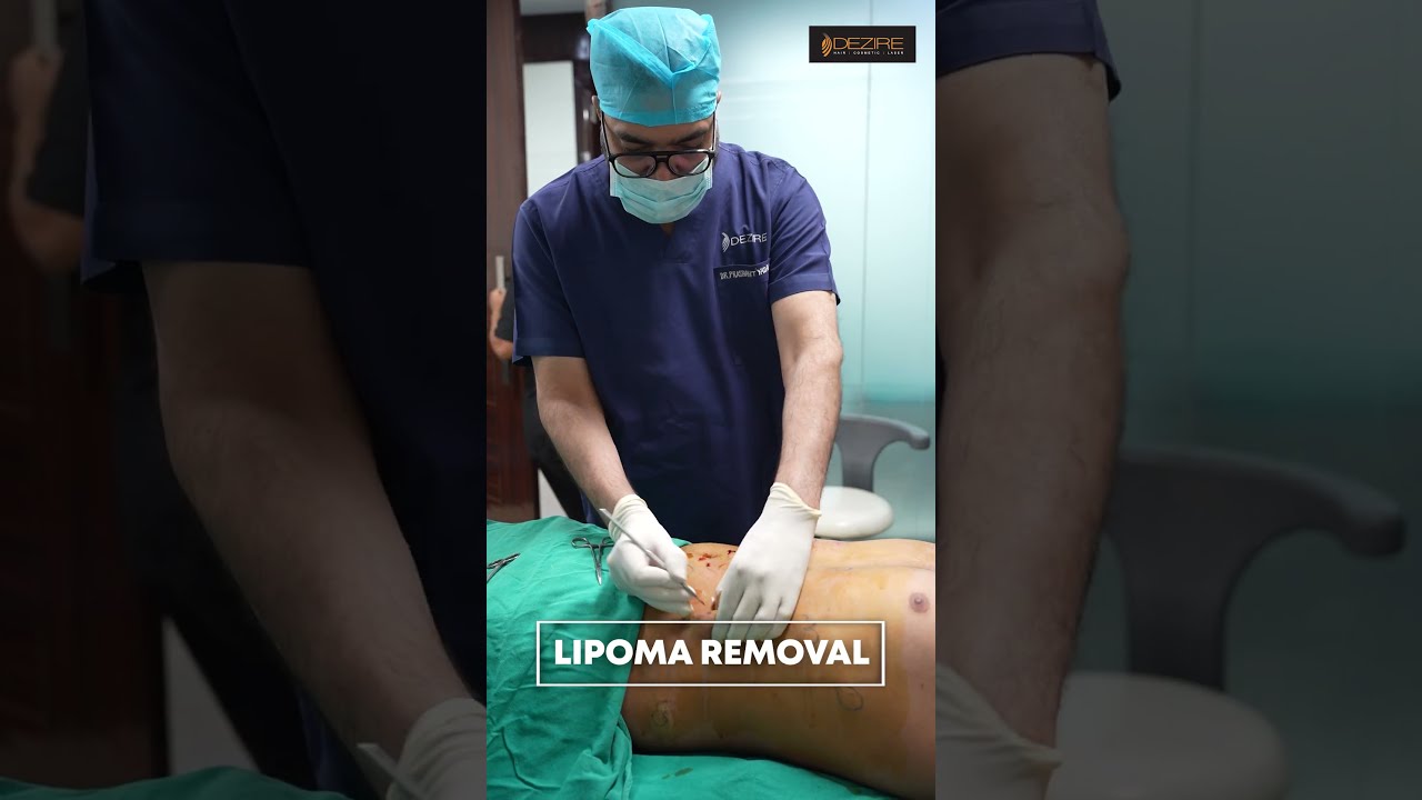
Image taken from the YouTube channel Dezire Clinic , from the video titled Lipoma cure | Multiple Lipoma removal #shorts .
Non-Encapsulated Lipoma Removal: Is It Possible? A Detailed Explanation
Lipomas are benign (non-cancerous) fatty tumors that grow slowly under the skin. They are typically encapsulated, meaning they are enclosed within a well-defined capsule of tissue. This encapsulation makes surgical removal relatively straightforward. However, when a lipoma is non-encapsulated – particularly when describing a non encapsulated lipoma with no clear boundaries removal – the challenge increases. Let’s explore this in detail.
Understanding Lipomas and Encapsulation
Before delving into removal, it’s crucial to understand the basic characteristics of lipomas.
-
What is a Lipoma? A lipoma is a soft, doughy mass composed of fat cells. They are usually painless and movable under the skin.
-
What Does "Encapsulated" Mean? Encapsulation refers to the presence of a distinct layer of fibrous tissue surrounding the lipoma. This capsule separates the lipoma from the surrounding tissues, making it easier to surgically dissect and remove. Think of it like peeling an orange; the peel is the capsule.
-
Why is Encapsulation Important for Removal? The capsule provides a clear plane of dissection, allowing the surgeon to cleanly remove the entire lipoma without disrupting surrounding tissues.
The Challenge of Non-Encapsulated Lipomas
The phrase "non encapsulated lipoma with no clear boundaries removal" highlights the core difficulty. When a lipoma lacks a capsule, it infiltrates the surrounding tissues, making its precise borders indistinct.
-
Infiltrative Growth: Non-encapsulated lipomas often grow in an infiltrative manner, meaning they extend finger-like projections into the muscle and subcutaneous fat.
-
Difficult Identification of Margins: Without a clear capsule, identifying the true extent of the lipoma becomes challenging. The surgeon may struggle to determine where the lipoma ends and healthy tissue begins.
-
Higher Risk of Incomplete Removal: Due to the ill-defined borders, there’s a higher risk of leaving behind remnants of the lipoma during surgery. This can lead to recurrence (the lipoma growing back).
Diagnostic Methods for Assessing Lipoma Characteristics
Accurate diagnosis and pre-operative assessment are crucial for planning the removal of a non-encapsulated lipoma with no clear boundaries.
-
Physical Examination: A careful physical examination can provide clues about the size, location, and consistency of the lipoma. Palpation (feeling the mass) can sometimes suggest the presence or absence of encapsulation.
-
Imaging Studies:
-
Ultrasound: Ultrasound is a non-invasive imaging technique that can help visualize the lipoma and assess its depth and relationship to surrounding structures.
-
MRI (Magnetic Resonance Imaging): MRI is generally considered the gold standard for evaluating lipomas, especially those suspected of being non-encapsulated. MRI provides detailed images of the lipoma and surrounding tissues, allowing the surgeon to better delineate its borders and identify any infiltrative growth.
-
CT Scan (Computed Tomography): CT scans can also be used, but they typically don’t provide the same level of detail as MRI for soft tissue masses like lipomas.
-
Example Table comparing Imaging Options:
Imaging Modality Advantages Disadvantages Usefulness for Non-Encapsulated Lipomas Ultrasound Non-invasive, inexpensive, readily available Limited detail, operator-dependent Initial assessment; less useful for deep, ill-defined lesions MRI High resolution, excellent soft tissue contrast, best for border definition More expensive, may not be available in all settings Highly recommended for suspected non-encapsulated lipomas CT Scan Good for overall anatomical assessment Less soft tissue detail than MRI, radiation exposure Less useful than MRI unless bony involvement is suspected
-
Surgical Techniques for Removing Non-Encapsulated Lipomas
The surgical approach for removing a non-encapsulated lipoma with no clear boundaries is more complex than for a typical, encapsulated lipoma.
-
Wide Excision: A wide excision involves removing the lipoma along with a margin of surrounding healthy tissue. This helps to ensure that all remnants of the lipoma are removed, reducing the risk of recurrence.
-
Liposuction: Liposuction may be used in conjunction with surgical excision, particularly for large lipomas or those with extensive infiltrative growth. Liposuction can help to remove some of the fatty tissue, making it easier to excise the remaining portion. However, liposuction alone is generally not recommended for non-encapsulated lipomas, as it may leave behind residual tumor cells.
-
En Bloc Resection: En bloc resection means removing the entire lipoma in one piece, without disrupting the tumor itself. This technique is often preferred for non-encapsulated lipomas to minimize the risk of spreading tumor cells.
-
Considerations for Deep Lipomas: When the lipoma is located deep within the muscle tissue, the surgeon may need to carefully dissect around important nerves and blood vessels. Nerve monitoring may be used during surgery to help prevent nerve damage.
-
Example Steps for Surgical Removal:
- Pre-operative Planning: Review imaging studies and plan the incision based on the location and extent of the lipoma.
- Incision and Exposure: Make an incision over the lipoma and carefully dissect through the skin and subcutaneous tissues to expose the tumor.
- Tumor Dissection: Identify the borders of the lipoma (which can be challenging in non-encapsulated cases). Carefully dissect around the lipoma, removing it along with a margin of surrounding healthy tissue.
- Hemostasis: Control any bleeding by cauterizing or ligating blood vessels.
- Closure: Close the incision in layers, using sutures to approximate the tissues.
Potential Risks and Complications
As with any surgical procedure, there are potential risks and complications associated with the removal of a non-encapsulated lipoma with no clear boundaries. These include:
- Recurrence: The most common risk is recurrence, especially if the lipoma is not completely removed.
- Seroma: A seroma is a collection of fluid under the skin. It can occur after surgery and may require drainage.
- Hematoma: A hematoma is a collection of blood under the skin. It can also occur after surgery and may require drainage.
- Infection: Infection is a risk with any surgical procedure. Antibiotics may be necessary to treat an infection.
- Nerve Damage: If the lipoma is located near a nerve, there is a risk of nerve damage during surgery. This can lead to numbness, tingling, or weakness in the affected area.
- Scarring: Scarring is inevitable after surgery. The appearance of the scar will depend on the size and location of the incision, as well as individual factors.
FAQs: Non-Encapsulated Lipoma Removal
Here are some common questions people have about removing non-encapsulated lipomas.
Can a non-encapsulated lipoma be removed?
Yes, it’s possible, but it’s generally more complex than removing an encapsulated lipoma. The key difference is that non-encapsulated lipomas blend into the surrounding tissue, making precise removal more challenging. The success depends on the lipoma’s size, location, and the surgeon’s skill.
What makes non-encapsulated lipoma removal difficult?
Because a non-encapsulated lipoma lacks a distinct capsule, it’s difficult to discern where the lipoma ends and healthy tissue begins. Removing all of the non encapsulated lipoma with no clear boundaries removal requires meticulous surgical technique to avoid damaging surrounding muscles, nerves, or blood vessels.
What are the treatment options for a non-encapsulated lipoma?
Surgical excision is often the primary treatment. Liposuction may also be considered, though complete removal may be harder to guarantee. In some cases, observation might be recommended if the lipoma is small, not causing symptoms, and the risks of removal outweigh the benefits. The choice depends on individual circumstances.
What are the risks associated with non-encapsulated lipoma removal?
Potential risks include incomplete removal (leading to recurrence), nerve damage, bleeding, scarring, and infection. Because of the ill-defined borders of a non encapsulated lipoma with no clear boundaries removal, the risk of these complications is generally higher compared to removing encapsulated lipomas. Discuss these risks with your surgeon.
So, there you have it! Hopefully, you’ve got a better handle on the complexities of non encapultated lipoma with no clear boundaries removal. If you’re facing this situation, remember to seek advice from a qualified professional. Good luck!