Mouse Chest Anatomy: Hidden Secrets Revealed! (60 Char)
The murine model serves as a cornerstone in biological research, offering critical insights into mammalian physiology. Understanding the precise anatomy mouse chest, therefore, becomes paramount for studies investigating respiratory mechanics, cardiovascular function, and immune responses. This intricate region, closely associated with the thoracic cavity, houses vital organs and complex musculoskeletal structures. Researchers at the Jackson Laboratory frequently employ advanced imaging techniques, such as micro-CT scanning, to meticulously analyze the anatomy mouse chest. Furthermore, a deep knowledge of the anatomy mouse chest is essential for scientists developing novel therapies targeting diseases affecting the cardiopulmonary system.
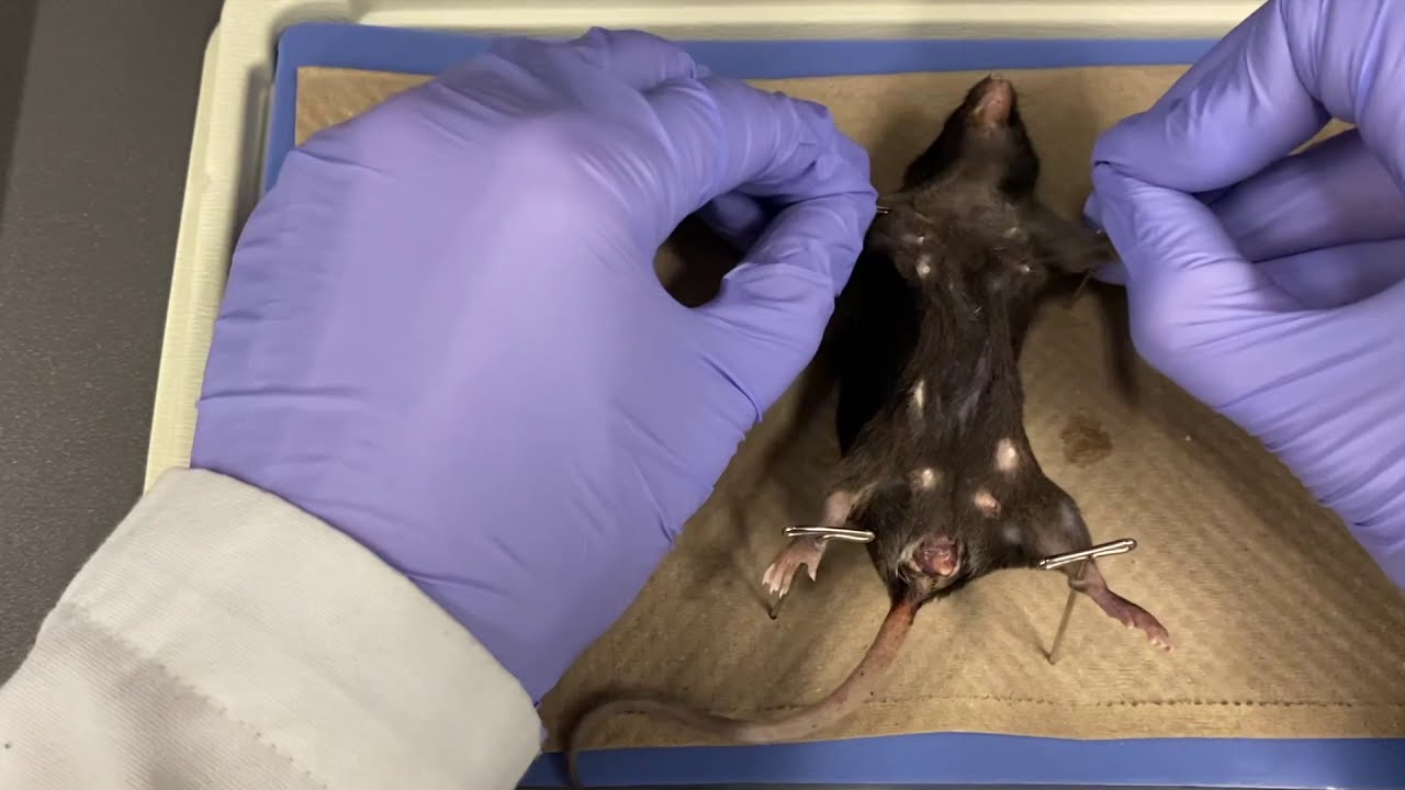
Image taken from the YouTube channel UTM Biology , from the video titled BIO153 Mouse Anatomy .
Mice are indispensable models in biomedical research, serving as critical surrogates for understanding human biology and disease. Their relatively short lifespans, genetic similarities to humans, and ease of manipulation make them ideal for studying a wide range of conditions, from cancer and cardiovascular disease to neurological disorders and infectious diseases. The insights gleaned from mouse studies are fundamental to developing new therapies and improving human health.
The Mouse as a Model Organism
The widespread use of mice in research underscores the critical need for a comprehensive understanding of their anatomy. Detailed anatomical knowledge is not merely academic; it is essential for accurate interpretation of experimental results, precise surgical interventions, and the development of targeted therapies.
However, delving into the anatomy of a mouse, particularly its thoracic cavity, reveals a world of intricate complexity.
The Hidden Complexity of the Mouse Chest
The mouse chest, or thoracic cavity, houses a delicate network of vital organs, including the heart, lungs, and major blood vessels. These structures are meticulously arranged and interconnected, working in concert to sustain life. The seemingly simple exterior of the mouse belies the sophisticated physiological processes occurring within its chest.
Understanding the spatial relationships between these organs, their microscopic structures, and their functional dynamics is crucial for researchers seeking to model human diseases accurately. Subtle variations in anatomy can significantly impact experimental outcomes, making detailed knowledge paramount.
Purpose and Scope
This editorial aims to provide a comprehensive overview of mouse chest anatomy, offering a detailed exploration of its bony structures, muscles, respiratory system, circulatory system, and supporting neurovascular elements. By elucidating the intricacies of the mouse thoracic cavity, this article seeks to empower researchers with the anatomical knowledge necessary to advance biomedical science and improve translational outcomes. It is intended to serve as a valuable resource for both seasoned investigators and newcomers to the field, fostering a deeper appreciation for the anatomical complexities of this vital model organism.
The mouse chest, as we’ve seen, is a marvel of biological engineering, a compact space housing essential organs. But before we can delve into the intricacies of the heart, lungs, and associated musculature, it’s crucial to understand the foundational framework that supports and protects these vital components. This bony architecture, comprised of the rib cage and sternum, provides the structural integrity necessary for respiration and shields the delicate organs from external forces.
The Foundation: Bony Structures of the Mouse Chest (Rib Cage and Sternum)
The skeletal framework of the mouse chest is a delicate yet robust structure, providing both protection and flexibility. It’s composed primarily of the rib cage and sternum, each playing a crucial role in the overall functionality of the thoracic cavity. Understanding the anatomy of these bony structures is fundamental to comprehending the biomechanics of respiration and the vulnerability of the internal organs.
The Rib Cage: A Protective Lattice
The rib cage in mice, like in most mammals, is a series of curved bones articulating with the vertebral column posteriorly and, in most cases, with the sternum anteriorly. However, there are notable differences in number and arrangement compared to humans.
Number and Arrangement of Ribs
Mice typically possess 13 pairs of ribs, a number that’s relatively consistent across different strains. These ribs can be categorized into true ribs, false ribs, and floating ribs, based on their anterior attachments.
The first seven ribs, termed true ribs, directly articulate with the sternum via costal cartilages. The subsequent five ribs are false ribs, indirectly connecting to the sternum through the costal cartilage of the rib above. The last rib pair are floating ribs or vertebral ribs, which do not articulate with the sternum at all, ending freely within the abdominal musculature. This arrangement provides flexibility to the rib cage during breathing and other movements.
Articulation with the Vertebral Column
Each rib articulates with the vertebral column at two points: the costovertebral joint (head of the rib with the vertebral body) and the costotransverse joint (tubercle of the rib with the transverse process of the vertebra).
These joints allow for a gliding motion, facilitating the expansion and contraction of the rib cage during respiration. The structural integrity of these articulations is crucial for maintaining the chest’s overall stability.
Protective Function
The primary function of the rib cage is to protect the vital organs within the thoracic cavity – the heart, lungs, and major blood vessels. The bony structure acts as a physical barrier against external trauma, minimizing the risk of injury to these delicate organs.
The curvature and elasticity of the ribs also contribute to their protective capacity, allowing them to absorb and distribute forces effectively. This protective function is paramount to the survival of the organism.
The Sternum: The Anchor Point
The sternum, or breastbone, is a flat, elongated bone located in the midline of the anterior chest wall. It serves as the anterior anchor point for the true and false ribs, contributing to the overall stability and structural integrity of the chest.
Structure and Components
The mouse sternum is comprised of multiple segments, though the exact number can vary slightly. These segments, known as sternebrae, are connected by cartilage, allowing for some degree of flexibility.
The most cranial segment, the manubrium, articulates with the clavicles (collarbones), while the remaining segments form the body of the sternum. The xiphoid process, the caudal-most segment, is typically cartilaginous.
Attachment Points for Ribs and Chest Muscles
The sternum provides attachment points for the costal cartilages of the true and false ribs, as well as for various chest muscles, including the pectoralis major and sternocleidomastoid. These attachments are critical for the proper function of the respiratory muscles and the overall biomechanics of the chest.
Role in Chest Stability and Structural Integrity
The sternum acts as a keystone, holding the rib cage together and providing stability to the entire thoracic structure. Its robust connection with the ribs ensures that the chest maintains its shape and resists deformation during respiration and other movements.
The sternum is crucial for maintaining the integrity of the chest cavity, protecting the organs within.
Contribution of the Musculoskeletal System
While the rib cage and sternum form the primary bony framework, it’s important to recognize the contribution of the broader musculoskeletal system within the chest cavity. The vertebral column, with its complex network of ligaments and muscles, provides posterior support and contributes to the overall flexibility of the chest.
The muscles of the chest wall, including the intercostals and diaphragm (discussed in the next section), interact directly with the bony structures, facilitating respiration and maintaining chest wall stability. The coordinated action of these musculoskeletal components is essential for proper chest function and overall health.
The skeletal framework of the mouse chest provides essential protection, but it’s the intricate network of muscles that truly brings the respiratory system to life. These muscles orchestrate the rhythmic expansion and contraction of the thoracic cavity, driving the vital process of gas exchange. Understanding their individual roles and coordinated actions is key to appreciating the elegant mechanics of mouse respiration.
Powering Respiration: Muscles of the Mouse Chest
The act of breathing, seemingly effortless, is in reality a complex interplay of various muscle groups within the thoracic cavity. In mice, as in other mammals, respiration is primarily driven by the coordinated action of the intercostal muscles and the diaphragm, each contributing uniquely to the process of inhalation and exhalation. The scalene muscles and serratus posterior muscles also play a role, albeit a less prominent one. A closer look at these muscle groups reveals the intricacies of respiratory mechanics in these vital research animals.
Muscles of Respiration: A Comprehensive Overview
The muscles of respiration in mice can be broadly categorized into those that primarily assist in inhalation and those involved in exhalation, although some muscles may contribute to both processes.
It is important to note that the contribution and activity level of each muscle can vary based on the physiological state of the mouse, such as during exercise or under anesthesia.
Intercostal Muscles: The Rib Cage Architects
The intercostal muscles, located between the ribs, are crucial in altering the volume of the thoracic cavity. They consist of two main layers: external and internal intercostals.
The external intercostal muscles run obliquely between the ribs, and their contraction elevates the rib cage, increasing the thoracic volume during inhalation.
In contrast, the internal intercostal muscles, with fibers running in an opposing direction, primarily contribute to exhalation by depressing the rib cage and decreasing thoracic volume.
Scalene Muscles: Neck Support, Respiratory Assist
The scalene muscles, located in the neck, also contribute to respiration by elevating the upper ribs. They provide additional support to the rib cage during deep or forced inhalation.
Their primary role is to assist in expanding the thoracic cavity when the mouse requires greater oxygen intake, such as during physical activity.
Serratus Posterior Superior and Inferior: Minor Players, Notable Roles
The serratus posterior superior and inferior muscles are thin, sheet-like muscles located on the posterior aspect of the thorax. The serratus posterior superior elevates the ribs, aiding in inhalation.
The serratus posterior inferior, on the other hand, depresses the ribs, assisting in exhalation.
While their contribution is less significant than that of the intercostals and diaphragm, they play a supporting role in respiration, particularly in maintaining chest wall stability.
Muscle Function During Inhalation and Exhalation
During inhalation, the diaphragm contracts and flattens, while the external intercostal and scalene muscles lift the rib cage. This coordinated action increases the volume of the thoracic cavity, creating a negative pressure that draws air into the lungs.
In contrast, exhalation is typically a passive process in mice at rest, resulting from the relaxation of the inspiratory muscles and the elastic recoil of the lungs and chest wall.
However, during forced exhalation, such as during coughing or sneezing, the internal intercostal muscles contract to actively reduce the thoracic volume and expel air from the lungs.
The Role of the Diaphragm: The Primary Driver of Respiration
The diaphragm, a large, dome-shaped muscle located at the base of the thoracic cavity, is the primary muscle of respiration in mice. Its contraction and relaxation drive the majority of air movement into and out of the lungs.
Origin, Insertion, and Innervation
The diaphragm originates from the xiphoid process of the sternum, the costal cartilages of the lower ribs, and the lumbar vertebrae. It inserts into a central tendon.
The phrenic nerve, originating from cervical spinal nerves, innervates the diaphragm, controlling its contraction and relaxation.
Mechanism of Diaphragmatic Breathing
During inhalation, the diaphragm contracts, pulling downward and flattening its dome shape. This increases the vertical dimension of the thoracic cavity, creating a negative pressure that draws air into the lungs.
During exhalation, the diaphragm relaxes, returning to its dome shape, which decreases the thoracic volume and forces air out of the lungs.
Importance in Respiratory Physiology
The diaphragm’s efficient and powerful contractions make it the most important muscle for normal breathing in mice. Damage or paralysis of the diaphragm can significantly impair respiratory function and compromise the animal’s ability to breathe.
Understanding the intricate interplay between the diaphragm and other respiratory muscles is critical for studying respiratory diseases and developing effective treatments.
Powering the respiratory mechanics with precisely orchestrated muscular movements is only part of the story. The ultimate destination of inhaled air, and the site of life-sustaining gas exchange, lies within the intricate architecture of the lungs and airways. Understanding the structure and function of these vital components is essential for a complete picture of mouse respiratory physiology.
The Respiratory Core: Lungs and Airways in Mice
The lungs and airways form the central hub of the respiratory system, responsible for the critical task of delivering oxygen to the bloodstream and removing carbon dioxide. In mice, as in other mammals, this system is exquisitely designed to maximize efficiency and ensure adequate gas exchange, even under varying physiological demands.
A detailed exploration of the murine lung reveals key adaptations and structural features that facilitate this vital process.
Lobar Architecture of the Mouse Lung
Unlike the human lung, which is divided into three lobes on the right and two on the left, the mouse lung exhibits a distinct lobar arrangement. The left lung consists of a single lobe, while the right lung is composed of four lobes: the cranial, middle, caudal, and accessory lobes.
This difference in lobation is noteworthy when comparing mouse models to human respiratory conditions.
The arrangement of these lobes within the thoracic cavity maximizes space utilization and ensures efficient ventilation of the entire lung volume. Each lobe is further subdivided into smaller units called lobules, creating a hierarchical branching pattern that increases the surface area available for gas exchange.
Microscopic Anatomy: Alveoli and Bronchioles
The true magic of respiration occurs at the microscopic level, within the alveoli and bronchioles. The bronchioles are tiny airways that branch off from the larger bronchi, ultimately leading to the alveoli. These structures represent the functional units of the lung, where oxygen and carbon dioxide are exchanged between the air and the blood.
Alveoli are tiny, sac-like structures with extremely thin walls, surrounded by a dense network of capillaries. This close proximity between air and blood facilitates the rapid diffusion of gases, driven by concentration gradients. The alveolar surface is also coated with a thin layer of surfactant, a substance that reduces surface tension and prevents the alveoli from collapsing.
The Gas Exchange Process
The gas exchange process within the lungs is a carefully orchestrated dance of diffusion and perfusion. Inhaled air, rich in oxygen, travels down the airways and into the alveoli. Here, oxygen diffuses across the alveolar-capillary membrane and into the bloodstream, where it binds to hemoglobin in red blood cells.
Simultaneously, carbon dioxide, a waste product of metabolism, diffuses from the blood into the alveoli and is exhaled from the body. The efficiency of this process depends on several factors, including the surface area of the alveoli, the thickness of the alveolar-capillary membrane, and the concentration gradients of oxygen and carbon dioxide.
Any disruption to these factors, such as in diseases like pneumonia or emphysema, can significantly impair gas exchange and lead to respiratory distress.
Integration with the Thoracic Cavity
The lungs and airways do not exist in isolation within the thoracic cavity. Their efficient function relies on their harmonious relationship with the other components of the chest, including the rib cage, muscles, heart, and blood vessels.
The rhythmic expansion and contraction of the rib cage, driven by the intercostal muscles and diaphragm, creates pressure gradients that draw air into and out of the lungs. The heart pumps blood through the pulmonary circulation, ensuring that the alveoli are adequately perfused with blood for gas exchange.
Nerves such as the vagus nerve also play a role by innervating the lungs and airways, controlling bronchoconstriction and mucus secretion. The intricate interplay between these structures highlights the complexity and elegance of the respiratory system, demonstrating how each component contributes to the overall goal of maintaining life-sustaining gas exchange.
The Circulatory System: Heart and Vessels of the Mouse Chest
The intricate orchestration of respiration hinges not only on muscular mechanics and pulmonary architecture, but also on the efficient delivery of oxygen and removal of carbon dioxide via the circulatory system. Within the mouse chest, the heart and major blood vessels form a dynamic network, seamlessly integrated with the respiratory system to sustain life. A thorough understanding of this cardiovascular component is crucial for interpreting physiological responses and modeling human diseases in murine studies.
A Detailed Examination of the Mouse Heart
The mouse heart, though small, shares fundamental structural similarities with the human heart, making it a valuable model for cardiovascular research. Understanding its precise location, internal anatomy, and function is paramount.
Precise Location within the Thoracic Cavity
The mouse heart resides within the mediastinum, the central compartment of the thoracic cavity. It is positioned slightly left of the midline, nestled between the lungs.
Its location is not arbitrary; the surrounding structures provide both support and protection, while also allowing for the necessary movement during contraction and relaxation.
Chambers, Valves, and Major Blood Vessels
The mouse heart, like the human heart, is a four-chambered organ, comprising two atria and two ventricles. The atria receive blood, while the ventricles pump blood out of the heart.
Valves ensure unidirectional blood flow: the tricuspid valve between the right atrium and right ventricle, the mitral valve (bicuspid) between the left atrium and left ventricle, the pulmonary valve between the right ventricle and pulmonary artery, and the aortic valve between the left ventricle and aorta.
Major blood vessels connected to the heart include the aorta, pulmonary artery, pulmonary veins, and vena cavae. These vessels are responsible for transporting blood to and from the lungs and the rest of the body.
The Cardiac Cycle and Blood Flow
The cardiac cycle describes the sequence of events that occur during one complete heartbeat. It consists of two main phases: systole (contraction) and diastole (relaxation).
During systole, the ventricles contract, pumping blood into the pulmonary artery (from the right ventricle) and the aorta (from the left ventricle). During diastole, the ventricles relax, allowing blood to flow from the atria into the ventricles.
Blood flow through the heart follows a specific pathway:
- Deoxygenated blood enters the right atrium via the vena cavae.
- Blood flows through the tricuspid valve into the right ventricle.
- The right ventricle pumps blood through the pulmonary valve into the pulmonary artery.
- The pulmonary artery carries blood to the lungs, where it becomes oxygenated.
- Oxygenated blood returns to the left atrium via the pulmonary veins.
- Blood flows through the mitral valve into the left ventricle.
- The left ventricle pumps blood through the aortic valve into the aorta.
- The aorta distributes oxygenated blood to the rest of the body.
Overview of Major Blood Vessels in the Chest
Beyond the heart itself, understanding the major blood vessels within the mouse chest is crucial for a complete picture of the circulatory system.
Aorta and its Significant Branches
The aorta is the largest artery in the body, originating from the left ventricle. Within the chest, the aorta gives rise to several significant branches that supply blood to the head, neck, and upper limbs. These branches include the brachiocephalic trunk, the left common carotid artery, and the left subclavian artery.
Pulmonary Artery and Veins
The pulmonary artery carries deoxygenated blood from the right ventricle to the lungs. It branches into the left and right pulmonary arteries, each supplying one lung. The pulmonary veins carry oxygenated blood from the lungs back to the left atrium of the heart.
Vena Cavae
The vena cavae are the largest veins in the body, responsible for returning deoxygenated blood from the body to the right atrium of the heart. The superior vena cava drains blood from the head, neck, and upper limbs, while the inferior vena cava drains blood from the trunk and lower limbs.
The Vital Role of the Cardiovascular System
The cardiovascular system plays an indispensable role in maintaining overall chest function and systemic homeostasis. Its primary function is to transport oxygen, nutrients, hormones, and immune cells to tissues throughout the body, while simultaneously removing carbon dioxide and waste products.
The heart’s efficient pumping action ensures continuous blood flow, while the intricate network of blood vessels facilitates the exchange of substances between the blood and surrounding tissues.
The cardiovascular system is intimately connected to the respiratory system, as the lungs are the site of gas exchange. The heart pumps deoxygenated blood to the lungs to pick up oxygen, and then pumps oxygenated blood to the rest of the body to deliver this vital gas. Disruptions to either system can have cascading effects on the other, highlighting the importance of understanding their interconnectedness.
Ultimately, a comprehensive understanding of the mouse heart and major blood vessels within the chest is essential for researchers seeking to model human cardiovascular diseases, test novel therapies, and unravel the complexities of cardiopulmonary physiology.
Supporting Structures: Nerves and Blood Vessels in the Mouse Chest
Beyond the major organs and skeletal framework, the mouse chest relies on a network of supporting structures: nerves and blood vessels. These often-overlooked components are critical for maintaining the health and functionality of the thoracic cavity. They ensure proper oxygenation, nutrient delivery, waste removal, and coordinated control of respiratory and cardiac functions.
Arterial Supply to the Mouse Chest
The arterial supply to the mouse chest is crucial for delivering oxygenated blood to the tissues and organs within the thoracic cavity.
Key arteries branch off the aorta to supply the chest wall, mediastinum, and lungs. Among the most important is the internal thoracic artery (also known as the internal mammary artery).
This artery courses along the inner surface of the chest wall, giving off branches that supply the intercostal spaces and contribute to the perfusion of the diaphragm.
Understanding the precise course and distribution of these arteries is essential in surgical procedures and when interpreting the effects of vascular diseases in murine models. Knowledge of typical artery branching patterns in mice is important when creating models of cardiovascular disease.
Venous Drainage of the Thoracic Region
The venous drainage system complements the arterial supply, removing deoxygenated blood and metabolic waste products from the chest.
Veins that drain the chest region, such as the internal thoracic veins (paralleling the arteries of the same name) and the azygos vein, play a significant role in this process.
The azygos vein, in particular, is a major vessel that collects blood from the intercostal veins and empties into the superior vena cava.
Efficient venous drainage is critical for preventing fluid accumulation and maintaining tissue homeostasis within the thoracic cavity. This drainage prevents fluid from impeding oxygen transfer.
Neural Control: The Phrenic and Vagus Nerves
The intricate functions of the mouse chest are under precise neural control, primarily mediated by the phrenic and vagus nerves.
The phrenic nerve is vital for diaphragmatic function, as it provides the primary motor innervation to the diaphragm.
Damage to the phrenic nerve can severely impair respiratory function, highlighting its importance in breathing.
The vagus nerve, on the other hand, has a more diverse role, influencing heart rate, bronchoconstriction, and gastrointestinal motility.
Its parasympathetic fibers innervate the lungs and heart, modulating their activity in response to various stimuli.
Dysfunction of the vagus nerve can lead to a range of respiratory and cardiovascular problems. Therefore, vagal nerve function is important to study in mouse models.
Maintaining Thoracic Health: An Integrated System
The nerves and blood vessels of the mouse chest act in concert to maintain the health and functionality of this vital region.
Arteries deliver oxygen and nutrients, veins remove waste products, and nerves coordinate muscle activity and organ function.
The integrity of these supporting structures is essential for ensuring proper respiration, circulation, and overall thoracic health.
Disruptions to this delicate balance can have significant consequences, underscoring the importance of understanding their anatomy and physiology in murine models of disease.
Studying nerves and blood vessels and their function in the mouse chest can inform a broad range of biomedical research.
Translational Significance: Clinical Relevance and Research Applications
The true value of understanding mouse chest anatomy extends far beyond mere academic curiosity.
A deep comprehension of the murine thorax unlocks critical insights and advancements across a spectrum of biomedical disciplines, from disease modeling to surgical innovation.
This section explores the multifaceted translational significance of murine thoracic anatomy, emphasizing its practical applications in addressing human health challenges.
Disease Modeling: Mimicking Human Ailments
Mice serve as indispensable models for studying human diseases due to their physiological similarities and genetic malleability.
A nuanced understanding of mouse chest anatomy is paramount for creating and interpreting these disease models, especially those concerning the respiratory and cardiovascular systems.
Respiratory Disease Modeling
Researchers leverage anatomical knowledge to induce and study conditions like asthma, chronic obstructive pulmonary disease (COPD), and pulmonary fibrosis in mice.
For instance, knowing the precise branching patterns of the murine airways is crucial for delivering targeted aerosolized drugs or inducing localized inflammation to mimic specific aspects of human lung diseases.
Furthermore, anatomical accuracy is vital for evaluating the efficacy of novel therapeutic interventions by assessing changes in lung structure and function.
Cardiovascular Disease Modeling
Similarly, mouse models are extensively used to investigate cardiovascular diseases such as myocardial infarction, atherosclerosis, and heart failure.
Understanding the intricate network of blood vessels supplying the heart and chest wall allows researchers to perform precise surgical procedures, such as coronary artery ligation, to simulate heart attacks.
Detailed anatomical knowledge also facilitates the accurate assessment of cardiac function and remodeling in response to various treatments.
Moreover, genetically modified mice with specific cardiovascular defects rely on careful anatomical phenotyping to fully characterize the disease phenotype.
Drug Development and Testing Protocols
The pharmaceutical industry relies heavily on preclinical studies in mice to evaluate the safety and efficacy of new drugs.
A thorough grasp of mouse chest anatomy is indispensable for designing and interpreting these studies, particularly for drugs targeting the respiratory or cardiovascular systems.
Optimizing Drug Delivery
The anatomical features of the mouse thorax influence the distribution and bioavailability of drugs administered via various routes, including inhalation, intravenous injection, or direct application to the chest wall.
Knowledge of the pulmonary vasculature and airway architecture is crucial for optimizing inhaled drug delivery to maximize therapeutic effects and minimize systemic exposure.
Similarly, understanding the anatomical relationships between blood vessels and surrounding tissues is essential for ensuring the accurate and targeted delivery of drugs to specific regions of the chest.
Assessing Drug Safety and Efficacy
During preclinical drug development, careful monitoring of the mouse chest is vital for detecting potential adverse effects, such as pulmonary edema, pleural effusion, or cardiac arrhythmias.
Anatomical knowledge is crucial for interpreting imaging data, such as chest X-rays or CT scans, to identify subtle structural changes indicative of drug-induced toxicity.
Furthermore, anatomical endpoints, such as lung volume, alveolar surface area, and cardiac output, are frequently used to assess the efficacy of drugs in preclinical studies.
Surgical Procedures and Interventions
Mouse models are increasingly used to develop and refine surgical techniques for treating chest diseases.
A comprehensive understanding of mouse chest anatomy is critical for performing these procedures safely and effectively.
Refining Surgical Techniques
Researchers use mouse models to practice and optimize surgical approaches for procedures such as lung resection, heart valve repair, and chest wall reconstruction.
Precise anatomical knowledge is essential for minimizing damage to surrounding tissues and ensuring successful outcomes.
For example, surgeons must be intimately familiar with the location of major blood vessels and nerves in the chest to avoid accidental injury during surgical procedures.
Developing Novel Interventions
Mouse models also play a crucial role in developing and testing new surgical interventions, such as minimally invasive techniques and implantable devices.
Anatomical considerations are paramount for designing these interventions and ensuring their compatibility with the unique anatomical features of the mouse chest.
For instance, the size and shape of implantable devices must be carefully tailored to the dimensions of the murine thorax to prevent complications such as compression of vital structures or migration of the device.
In conclusion, the translational significance of understanding mouse chest anatomy cannot be overstated.
Its applications span a wide range of biomedical disciplines, from disease modeling and drug development to surgical innovation.
By leveraging our knowledge of the murine thorax, we can advance our understanding of human diseases and develop more effective treatments for a variety of life-threatening conditions.
Mouse Chest Anatomy FAQs
Here are some frequently asked questions about the fascinating hidden secrets of the mouse chest anatomy, offering clarity and deeper insights into this vital area.
What are the major components within the mouse chest cavity?
The mouse chest cavity, like that of other mammals, houses crucial organs including the heart, lungs, esophagus, and trachea. Understanding the anatomy mouse chest requires knowing these components work together. Major blood vessels also traverse this space.
How does the rib cage protect the internal organs in the mouse chest?
The rib cage provides a bony shield safeguarding the delicate organs within the mouse chest. This protection is essential for survival, shielding against external impact and injury. The rib cage allows for respiratory movement.
Is the heart positioned differently in a mouse compared to a human?
While the basic function is the same, the relative position of the heart within the anatomy mouse chest can differ slightly from humans due to body size and overall structure. It’s proportionally larger in mice.
What is the role of the diaphragm in mouse chest respiration?
The diaphragm, a muscle separating the chest and abdominal cavities, plays a key role in mouse chest respiration. Its contraction and relaxation generate pressure changes, enabling efficient breathing and ventilation of the lungs. Its movement influences airflow.
So, that’s a wrap on the anatomy mouse chest! Hopefully, you found this dive into murine thoracic architecture enlightening. Now you’re practically an expert on the subject! Keep exploring and learning!
