Mitosis & Meiosis: Master the Index! [Easy Guide]
Cell division, a fundamental process explored through microscopy, underpins growth and reproduction. Mitosis and meiosis, two distinct types of cell division, are central to understanding this biological phenomenon. One key technique, extensively used in cancer research, involves observing mitosis and determining meiotic index to assess cell proliferation rates. The protocol developed by Dr. Sharma’s lab outlines effective methods for sample preparation and data analysis, while ImageJ provides powerful tools for quantifying cellular activity and accurately observing mitosis and determining meiotic index, furthering our understanding of cell cycle regulation.
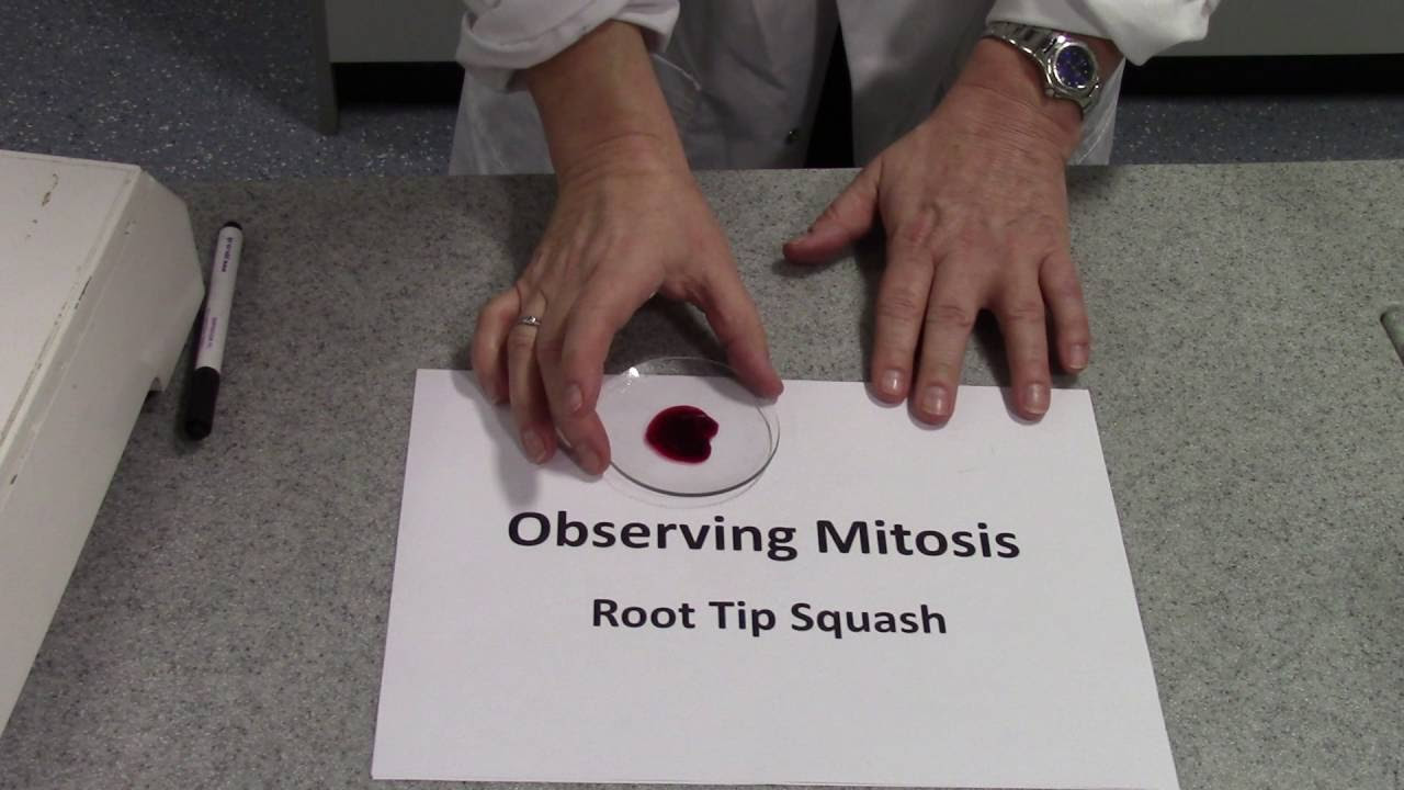
Image taken from the YouTube channel Biology Practicals and Revision Biology Tutor , from the video titled Mitotic Index Root Tip Squash .
Mastering Mitosis & Meiosis: A Guide to Observation and Indexing
This guide provides a structured approach to understanding and observing mitosis and meiosis, focusing on calculating the meiotic index. It aims to break down complex biological processes into manageable steps for easier comprehension and application.
Understanding Mitosis and Meiosis
Before diving into observation and indexing, it’s crucial to establish a clear understanding of what mitosis and meiosis are.
Mitosis: Cell Replication for Growth and Repair
Mitosis is the process of cell division that results in two identical daughter cells, each having the same number and kind of chromosomes as the parent nucleus. It’s essential for growth, repair, and asexual reproduction.
- Key Stages:
- Prophase: Chromosomes condense and become visible. The nuclear envelope breaks down.
- Metaphase: Chromosomes align along the metaphase plate (equator) of the cell.
- Anaphase: Sister chromatids separate and move to opposite poles of the cell.
- Telophase: Chromosomes arrive at the poles and begin to decondense. The nuclear envelope reforms.
- Cytokinesis: The cytoplasm divides, resulting in two separate daughter cells.
Meiosis: Producing Genetically Diverse Gametes
Meiosis is a specialized type of cell division that reduces the chromosome number by half, resulting in four genetically different daughter cells. It’s vital for sexual reproduction, ensuring genetic diversity in offspring.
- Key Stages (Meiosis I and Meiosis II):
- Meiosis I:
- Prophase I: Chromosomes condense, homologous chromosomes pair up (synapsis), and crossing over occurs (exchange of genetic material).
- Metaphase I: Homologous chromosome pairs align along the metaphase plate.
- Anaphase I: Homologous chromosomes separate and move to opposite poles (sister chromatids remain attached).
- Telophase I: Chromosomes arrive at the poles. Cytokinesis usually occurs, resulting in two haploid cells.
- Meiosis II: Similar to mitosis, but with haploid cells.
- Prophase II: Chromosomes condense again.
- Metaphase II: Chromosomes align along the metaphase plate.
- Anaphase II: Sister chromatids separate and move to opposite poles.
- Telophase II: Chromosomes arrive at the poles. Cytokinesis occurs, resulting in four haploid daughter cells (gametes).
- Meiosis I:
Observing Mitosis and Meiosis
Effective observation relies on proper preparation and clear identification of cellular structures.
Mitosis Observation Techniques
-
Microscopy: Utilize a microscope with sufficient magnification (typically 400x or higher) to visualize chromosomes and cellular structures.
-
Sample Preparation: Plant root tips (e.g., onion root tips) are commonly used due to their actively dividing cells.
- Fixation: Preserve the tissue structure using a fixative (e.g., acetic acid and ethanol).
- Staining: Enhance visibility of chromosomes using stains (e.g., acetocarmine or Feulgen stain).
- Squashing: Gently squash the tissue on a slide to create a single layer of cells.
-
Identifying Mitotic Stages: Carefully observe the distinct characteristics of each stage (prophase, metaphase, anaphase, telophase) based on chromosome behavior and cell structure.
Meiosis Observation Techniques
- Microscopy: As with mitosis, a microscope with high magnification is necessary.
- Sample Preparation: Focus on reproductive organs (e.g., anthers in plants or testes in animals) where meiosis occurs.
- Fixation, Staining, and Squashing: Similar preparation methods are used as with mitosis, but may require specialized staining techniques to highlight synaptonemal complexes or chiasmata (points of crossing over).
- Identifying Meiotic Stages: This requires meticulous observation due to the complexity of the process. Look for:
- Synapsis and crossing over in Prophase I.
- Alignment of homologous chromosomes in Metaphase I.
- Separation of homologous chromosomes in Anaphase I.
- Separation of sister chromatids in Anaphase II.
Determining the Meiotic Index
The meiotic index provides a quantitative measure of meiotic activity in a tissue sample. It is calculated as the percentage of cells undergoing meiosis.
Calculating the Meiotic Index: A Step-by-Step Guide
- Sample Selection: Choose representative fields of view under the microscope.
- Cell Counting: Count the total number of cells in each field of view. This should include cells in all stages of the cell cycle, as well as those not actively dividing.
- Meiotic Cell Count: Identify and count the number of cells exhibiting clear meiotic stages (Prophase I, Metaphase I, Anaphase I, Telophase I, Prophase II, Metaphase II, Anaphase II, Telophase II).
-
Index Calculation: Calculate the meiotic index using the following formula:
Meiotic Index = (Number of Meiotic Cells / Total Number of Cells) x 100Express the result as a percentage.
Factors Affecting Meiotic Index
Several factors can influence the meiotic index, including:
- Tissue Type: Different tissues have varying levels of meiotic activity.
- Developmental Stage: Meiotic activity can fluctuate during development.
- Environmental Factors: Stress or exposure to certain chemicals can affect meiosis.
- Sampling Technique: Appropriate sampling is crucial for obtaining accurate and representative data.
Example Calculation
Let’s say you examine a slide of an anther and count the following in one field of view:
- Total Number of Cells: 200
- Number of Meiotic Cells: 30
The meiotic index would be calculated as follows:
Meiotic Index = (30 / 200) x 100 = 15%
This indicates that 15% of the cells in that field of view are undergoing meiosis. Repeat this calculation across multiple fields of view and average the results for a more accurate representation of the overall meiotic index.
Mitosis & Meiosis FAQ: Your Questions Answered
This FAQ addresses common questions about mitosis and meiosis to help you master the index and understand these critical cell division processes.
What’s the key difference between mitosis and meiosis?
Mitosis results in two identical daughter cells, used for growth and repair. Meiosis, however, produces four genetically distinct daughter cells, essential for sexual reproduction. Observing mitosis and determining meiotic index are very different processes.
Why is the meiotic index important?
The meiotic index helps assess the rate of cell division during meiosis in a sample. This is crucial in studies of fertility, plant breeding, and understanding the effects of certain chemicals on reproductive cells.
What are the main phases of mitosis, in order?
The phases of mitosis are: Prophase, Metaphase, Anaphase, and Telophase (PMAT). Remember these phases to understand cell division! Studying these phases aids in observing mitosis.
Where does meiosis typically occur?
Meiosis occurs in the reproductive organs: in animals, it’s the testes and ovaries; in plants, it’s the anthers and ovaries. Studying these parts helps with observing mitosis and determining meiotic index.
And there you have it! Hopefully, you feel a little more confident now about observing mitosis and determining meiotic index. Go ahead and put those newfound skills to work – good luck!
