Lamina Dura & Bone Anatomy: Vital Facts You Need To Know
Understanding the lamina dura / compact bone head and necl anatomy is foundational in dentistry and bone physiology. Radiographic interpretation heavily relies on the assessment of the lamina dura’s integrity, as changes in its appearance often signal underlying dental or systemic conditions. The mandible, a critical structure discussed frequently in the context of oral surgery, exhibits unique bone density patterns related to the lamina dura and the trabecular bone supporting teeth. Further, the biomechanical properties of the compact bone are often studied using finite element analysis to model stress distribution around dental implants anchored within the alveolar bone.
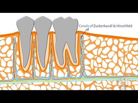
Image taken from the YouTube channel Doctoropsy , from the video titled Alveolar Bone- Structure .
Structuring an Article on Lamina Dura & Bone Anatomy
This outline details an effective article structure focusing on the lamina dura and related bone anatomy, targeting the keyword "lamina dura / compact bone head and neck anatomy."
I. Introduction: Setting the Stage
- Start with a brief, engaging overview of bone health and its importance. Hint at the crucial role the lamina dura plays.
- Immediately introduce the lamina dura and its location in the context of teeth and alveolar bone.
- Mention the significance of understanding the lamina dura for dental professionals and anyone interested in oral health.
- Clearly state the article’s objective: to provide a comprehensive overview of the lamina dura and related bone structures, especially concerning the compact bone in the head and neck.
II. Defining the Lamina Dura
A. What is the Lamina Dura?
- Provide a detailed definition of the lamina dura. Emphasize that it’s the bone lining the tooth socket (alveolus).
- Explain its composition: dense, compact bone (specifically, bundle bone) rich in Sharpey’s fibers. These are collagen fibers from the periodontal ligament (PDL) that insert into the bone.
- Use descriptive language: "a thin, radiopaque (appearing white on X-rays) layer…"
- Mention its continuous nature with the surrounding alveolar bone.
B. Function of the Lamina Dura
- Explain the primary function: attachment point for the periodontal ligament fibers.
- Highlight its role in anchoring the tooth within the alveolar socket.
- Describe its involvement in the tooth’s response to occlusal forces (biting). The lamina dura helps distribute and withstand these forces.
- Emphasize its dynamic nature – it can remodel and adapt in response to changes in forces applied to the tooth.
III. Compact Bone Anatomy in the Head and Neck: A Broader View
A. Overview of Bone Types
- Briefly describe the two main types of bone tissue: compact (cortical) bone and spongy (cancellous/trabecular) bone.
- Focus on compact bone, highlighting its density and strength.
- Explain the location of compact bone: the outer layer of most bones, including those in the head and neck.
B. Detailed Structure of Compact Bone
- Haversian Systems (Osteons):
- Describe the osteon as the fundamental functional unit of compact bone.
- Explain the concentric arrangement of lamellae (layers of bone matrix) around a central Haversian canal.
- Describe the Haversian canal’s contents: blood vessels, nerves, and lymphatic vessels.
- Lacunae and Canaliculi:
- Explain that osteocytes (bone cells) reside within lacunae (small cavities) between lamellae.
- Describe canaliculi: tiny channels radiating from lacunae, allowing communication and nutrient exchange between osteocytes.
- Volkmann’s Canals (Perforating Canals):
- Describe Volkmann’s canals as channels that connect Haversian canals, allowing blood vessels and nerves to travel between osteons and to the bone surface.
C. Compact Bone in Specific Head and Neck Bones
- Mandible (Lower Jaw):
- Describe the thick layer of compact bone forming the outer surface of the mandible.
- Explain its role in protecting the underlying spongy bone and housing the tooth sockets.
- Maxilla (Upper Jaw):
- Describe the compact bone structure of the maxilla, noting its relatively thinner layer compared to the mandible in some areas.
- Explain its contribution to the hard palate and nasal floor.
- Cranial Bones:
- Briefly mention the role of compact bone in the cranial bones, such as the frontal, parietal, temporal, and occipital bones, forming the skull and protecting the brain. Include a brief overview of the inner and outer tables.
IV. The Lamina Dura and its Radiographic Appearance
A. How the Lamina Dura Appears on Dental X-rays
- Explain that the lamina dura appears as a distinct, radiopaque (white) line surrounding the tooth root on radiographs.
- Describe the normal appearance: uniform thickness and continuity.
B. Variations in Appearance
- Thickening:
- Discuss potential causes of a thickened lamina dura: increased occlusal forces (bruxism), orthodontic treatment.
- Thinning or Loss:
- Discuss potential causes of a thinned or absent lamina dura: periodontal disease, periapical lesions, certain systemic conditions (e.g., osteoporosis).
- Factors Affecting Radiographic Interpretation:
- Mention factors that can affect the appearance of the lamina dura on radiographs: angulation of the X-ray beam, quality of the radiograph.
V. Clinical Significance: Lamina Dura as a Diagnostic Indicator
A. Importance in Diagnosing Periodontal Disease
- Explain how changes in the lamina dura can indicate the presence and severity of periodontal disease.
- Describe how bone loss (resorption) around the tooth, evidenced by a loss of lamina dura definition, is a key sign of periodontal disease progression.
B. Importance in Identifying Periapical Lesions
- Explain how periapical lesions (infections around the tooth root) can cause destruction of the lamina dura in the apical region.
- Describe how a break in the lamina dura continuity near the apex can indicate a periapical abscess or granuloma.
C. Role in Orthodontic Treatment
- Explain that orthodontic treatment (braces) relies on bone remodeling around the teeth.
- Describe how the lamina dura changes during orthodontic treatment as teeth move through the bone. The lamina dura resorbs on the pressure side and forms on the tension side.
D. The Lamina Dura and Systemic Diseases
- Mention that certain systemic diseases, such as osteoporosis, can affect bone density and, consequently, the appearance of the lamina dura.
VI. Maintaining a Healthy Lamina Dura
- Oral Hygiene:
- Emphasize the importance of good oral hygiene practices (brushing, flossing) to prevent periodontal disease and maintain a healthy lamina dura.
- Regular Dental Checkups:
- Stress the need for regular dental checkups and radiographs to monitor the health of the lamina dura and detect any early signs of problems.
- Balanced Diet:
- Explain the importance of a balanced diet rich in calcium and vitamin D for maintaining healthy bone density, including the lamina dura.
- Avoiding Parafunctional Habits:
- Advise patients to avoid habits like teeth grinding (bruxism) that can put excessive stress on the lamina dura.
Frequently Asked Questions: Lamina Dura & Bone Anatomy
Here are some frequently asked questions to help clarify vital facts about the lamina dura and related bone anatomy.
What exactly is the lamina dura?
The lamina dura is a thin, radiopaque (appears white on X-rays) layer of dense compact bone that surrounds the tooth socket. It’s part of the alveolar bone, which supports the teeth. Think of it as the inner wall of the bony housing for your tooth root.
How does the lamina dura relate to overall bone health?
The lamina dura’s appearance on radiographs can be an indicator of bone metabolic activity. Changes in its density or thickness can suggest underlying bone diseases or conditions affecting compact bone head and neck anatomy, as well as metabolic processes affecting the skeletal system.
What’s the clinical significance of the lamina dura?
Dentists and radiologists assess the lamina dura on dental X-rays to evaluate periodontal health and detect abnormalities. A loss or thinning of the lamina dura can suggest conditions like periodontitis, hyperparathyroidism, or even certain types of bone cancers.
How does compact bone contribute to the overall structure of the skull?
Compact bone forms the outer layer of most bones, including those in the skull. It provides strength and protection. The structure of the lamina dura, being specialized compact bone, demonstrates how localized bone density variations support specific functions within the skeletal framework, such as securely anchoring teeth within the compact bone head and neck anatomy.
So, there you have it – a quick rundown on the key aspects of lamina dura / compact bone head and necl anatomy! Hopefully, this helps you better understand its importance. Until next time!
