Heart Anatomy: The Ultimate Visual Guide!
Understanding the circulatory system requires a deep dive into the anatomy of the heart (e.g., its intricate structure and function. Medical students frequently utilize resources like the Gray’s Anatomy textbook to study the heart’s chambers and valves. The American Heart Association plays a vital role in disseminating information about cardiovascular health, including details on the anatomy of the heart (e.g.. Visual learning tools, such as 3D heart models, enhance comprehension of the spatial relationships within the organ, enabling a better grasp of the anatomy of the heart (e.g.. Cardiac surgeons, practicing in advanced medical centers, possess expert knowledge of the anatomy of the heart (e.g., allowing them to perform complex procedures to repair or replace damaged heart tissues.
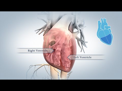
Image taken from the YouTube channel Nucleus Medical Media , from the video titled Anatomy of the Heart .
Heart Anatomy: Crafting the Ultimate Visual Guide Article
To create an engaging and informative "Heart Anatomy: The Ultimate Visual Guide!" article, a structured and visually rich layout is crucial. The primary focus must be on "the anatomy of the heart," making it clear, accessible, and memorable for readers of all backgrounds.
I. Introduction: Setting the Stage for Understanding
The introduction should immediately grab the reader’s attention and clearly define the purpose of the article.
- Engaging Hook: Start with a captivating fact or statistic about the heart’s importance. Example: "Did you know your heart beats around 100,000 times a day, pumping life-giving blood throughout your entire body? Understanding how this incredible organ works starts with understanding its anatomy."
- Brief Overview: Concisely explain what the article will cover, focusing on key anatomical structures and their functions.
- Relevance & Benefits: Highlight why understanding heart anatomy is essential for general knowledge, health awareness, and potentially for those studying related fields.
- Visual Cue: Ideally, feature a compelling introductory image or illustration of the heart.
II. External Anatomy: A Bird’s-Eye View
This section focuses on the heart’s external appearance and features.
A. Size and Shape
- Description: Describe the heart’s approximate size and shape (e.g., "about the size of a fist," cone-shaped).
- Visuals: Include illustrations or photographs showcasing the heart’s overall form and dimensions. Use labels to identify major features.
- Orientation: Explain the heart’s position within the chest cavity (mediastinum) and its orientation (apex pointing downwards and to the left).
B. Surface Features
- Description: Detail the visible features on the heart’s surface, such as:
- Atria: The upper chambers.
- Ventricles: The lower chambers.
- Sulci (Grooves): Coronary sulcus (atrioventricular groove) and interventricular sulcus. Explain their locations and importance (containing coronary arteries and veins).
- Visuals: Use annotated diagrams or labeled photographs to clearly identify these surface features. Consider using different colors for better differentiation.
C. Major Vessels
- Description: Identify and describe the major blood vessels connected to the heart:
- Aorta: Carries oxygenated blood to the body.
- Pulmonary Artery: Carries deoxygenated blood to the lungs.
- Superior and Inferior Vena Cava: Carry deoxygenated blood from the body to the heart.
- Pulmonary Veins: Carry oxygenated blood from the lungs to the heart.
- Visuals: Emphasize these vessels in the visual aids, highlighting their points of connection to the heart. Color-coding can be very effective here (e.g., red for arteries, blue for veins).
III. Internal Anatomy: Peering Inside the Heart
This section delves into the inner workings and components of the heart.
A. Chambers
- Description: Describe each chamber in detail:
- Right Atrium: Receives deoxygenated blood.
- Right Ventricle: Pumps deoxygenated blood to the lungs.
- Left Atrium: Receives oxygenated blood.
- Left Ventricle: Pumps oxygenated blood to the body.
- Visuals: Utilize cross-sectional diagrams or 3D renderings of the heart to reveal the internal chambers. Label each chamber clearly.
B. Valves
- Description: Explain the function and structure of the four heart valves:
- Tricuspid Valve: Between the right atrium and right ventricle.
- Pulmonary Valve: Between the right ventricle and pulmonary artery.
- Mitral Valve (Bicuspid): Between the left atrium and left ventricle.
- Aortic Valve: Between the left ventricle and aorta.
- Visuals: Include close-up illustrations or diagrams of each valve, showing the leaflets (cusps) and their mechanisms of opening and closing. Animated GIFs can be extremely effective for demonstrating valve function.
-
Table: Create a table summarizing the valves:
Valve Name Location Function Tricuspid Right Atrium to Right Ventricle Prevents backflow of blood from the right ventricle to the right atrium. Pulmonary Right Ventricle to Pulmonary Artery Prevents backflow of blood from the pulmonary artery to the right ventricle. Mitral (Bicuspid) Left Atrium to Left Ventricle Prevents backflow of blood from the left ventricle to the left atrium. Aortic Left Ventricle to Aorta Prevents backflow of blood from the aorta to the left ventricle.
C. Walls
- Description: Describe the layers of the heart wall:
- Epicardium: Outer layer.
- Myocardium: Muscular middle layer (responsible for contraction).
- Endocardium: Inner lining.
- Visuals: Use a detailed cross-sectional diagram of the heart wall to illustrate these layers. Emphasize the thickness of the myocardium, particularly in the left ventricle.
D. Septa
- Description: Explain the septa (walls) that divide the heart:
- Interatrial Septum: Separates the atria.
- Interventricular Septum: Separates the ventricles.
- Visuals: Clearly indicate the septa in the internal heart diagrams.
E. Chordae Tendineae and Papillary Muscles
- Description: Explain the function of chordae tendineae (heart strings) and papillary muscles in preventing valve prolapse (backflow of blood).
- Visuals: Show how these structures connect to the valves in the illustrations.
IV. Blood Supply: Nourishing the Heart
This section explains how the heart muscle receives its own blood supply.
A. Coronary Arteries
- Description: Detail the origins, paths, and branches of the major coronary arteries (left and right coronary arteries). Explain how these arteries supply oxygenated blood to the myocardium.
- Visuals: Use a diagram illustrating the coronary arteries as they wrap around the heart. Color-code the arteries (e.g., red) to differentiate them clearly. Indicate major branches like the left anterior descending (LAD) artery and the circumflex artery.
B. Coronary Veins
- Description: Explain how deoxygenated blood is drained from the myocardium by the coronary veins, primarily draining into the coronary sinus.
- Visuals: Show the coronary veins in conjunction with the coronary arteries, using a different color (e.g., blue) to distinguish them.
V. Nerve Supply: Regulating Heart Rate
This section describes the heart’s intrinsic conduction system and autonomic nervous system influence.
A. Intrinsic Conduction System
- Description: Explain the components and function of the heart’s internal electrical system:
- Sinoatrial (SA) Node (Pacemaker): Initiates the heartbeat.
- Atrioventricular (AV) Node: Delays the signal.
- Bundle of His: Transmits the signal to the ventricles.
- Purkinje Fibers: Distribute the signal throughout the ventricular myocardium.
- Visuals: Use a diagram showing the conduction pathway through the heart, with arrows indicating the direction of electrical impulses.
B. Autonomic Innervation
- Description: Briefly mention how the sympathetic and parasympathetic nervous systems influence heart rate and contractility.
- Visuals: An overview illustration can suffice for this section, showing the sympathetic and parasympathetic nerves connecting to the heart.
By following this detailed layout, the article "Heart Anatomy: The Ultimate Visual Guide!" will offer a comprehensive and visually engaging exploration of the anatomy of the heart.
FAQs About Heart Anatomy
Here are some frequently asked questions regarding the anatomy of the heart, to help clarify some key concepts covered in our visual guide.
What are the four chambers of the heart and what are their primary functions?
The heart has four chambers: the left and right atria, and the left and right ventricles. The atria receive blood, while the ventricles pump blood out of the heart. Understanding the anatomy of the heart (e.g., how blood flows through these chambers) is crucial for grasping its overall function.
How do the valves of the heart work and why are they important?
Heart valves (tricuspid, mitral, pulmonary, and aortic) ensure blood flows in one direction. They open and close in response to pressure changes within the chambers. Proper valve function is vital to the anatomy of the heart. Dysfunction can lead to heart failure.
What is the coronary circulation, and why is it so critical?
The coronary circulation provides blood supply to the heart muscle itself. Blockage of coronary arteries can lead to a heart attack. This system shows a critical aspect of the anatomy of the heart.
Can you explain the difference between the pulmonary and systemic circuits?
The pulmonary circuit carries blood from the heart to the lungs for oxygenation and back. The systemic circuit carries oxygenated blood from the heart to the rest of the body and returns deoxygenated blood back. These circuits showcase a key aspect of the anatomy of the heart and blood flow through it.
So there you have it – your ultimate visual guide to the anatomy of the heart (e.g.! Hope you found it helpful. Keep on exploring, and take good care of your ticker!
