Bony Flex Explained: Is it the Key to Ankle Health?
Ankle health, a cornerstone of overall mobility, hinges on complex biomechanics. The lateral malleolus, a distal fibula landmark, serves as a crucial reference point when assessing ankle joint stability. Understanding the bony fkex inferior to lateral malleolus, as it relates to proprioception and ankle functionality, has become increasingly important for practitioners in sports medicine. Its proper assessment is foundational for accurate diagnoses.
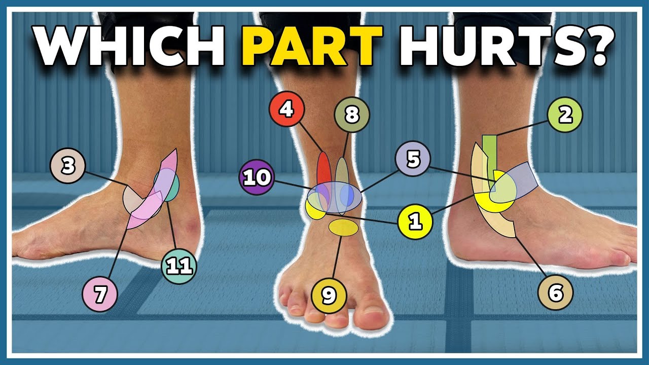
Image taken from the YouTube channel Prof. Dr. J. Bellemans , from the video titled My ankle hurts here! 11 typical pain spots and what they mean .
Unlocking Ankle Health Through the Bony Flex
The ankle, a seemingly simple joint, is a complex structure crucial for countless daily activities. From walking and running to simply maintaining balance, healthy ankles are essential for mobility and overall well-being. When ankle function is compromised, it can have a ripple effect, impacting everything from athletic performance to everyday tasks.
At the heart of optimal ankle function lies a subtle yet significant component often overlooked: the bony flex. This refers to the slight give or movement that occurs at a specific point on the outside of the ankle. Understanding the bony flex is key to preventing injuries and maintaining long-term ankle health.
The Bony Flex: A Key Player in Ankle Mechanics
The bony flex is intimately connected to the overall function of the ankle joint. It’s a subtle movement, not a dramatic bend or twist, but rather a slight yielding that accommodates the complex mechanics of the ankle during motion.
Think of it as a shock absorber or a fine-tuning mechanism that allows the ankle to adapt to uneven surfaces and varying loads.
Its presence indicates healthy joint mobility and proper alignment of the surrounding structures. A restricted bony flex, on the other hand, can signal underlying problems that may lead to pain and injury.
Pinpointing the Location: Inferior to the Lateral Malleolus
To understand the bony flex, it’s essential to know its location. It can be found just below the lateral malleolus, which is the bony prominence on the outer side of your ankle. This is the end of the fibula bone, and it plays a critical role in ankle stability.
The bony flex isn’t the lateral malleolus itself, but rather the area immediately inferior to it. Palpating this area, you may feel a subtle "give" or springiness when the ankle is moved gently. This sensation indicates a healthy and functional bony flex.
Exploring the Bony Flex: Our Goal
This article aims to define, explain, and emphasize the importance of the bony flex in maintaining ankle health.
We will explore the anatomy involved, delve into the biomechanics of this subtle movement, and discuss its critical role in preventing injuries.
By the end of this exploration, you’ll gain a deeper understanding of your ankle’s inner workings and how to prioritize its long-term well-being.
Ankle Anatomy 101: Key Structures and Their Roles
Before diving deeper into the mechanics of the bony flex, it’s crucial to establish a solid foundation in ankle anatomy. This section provides a concise yet comprehensive overview of the key anatomical structures directly involved in this subtle yet significant movement. Understanding these structures is essential for grasping the biomechanics and overall importance of the bony flex in maintaining a healthy, functional ankle.
The Bony Players: A Foundation of Support
The ankle joint, or talocrural joint, is a complex articulation formed by the tibia, fibula, and talus. The tibia and fibula, the two long bones of the lower leg, come together to create a mortise, a socket that cradles the talus, the uppermost bone of the foot. Understanding the roles of these bones is paramount to understanding ankle function.
Lateral Malleolus: The Fibula’s Distal Extension
The lateral malleolus is the prominent bony projection on the outer side of your ankle. This is the distal (lower) end of the fibula. Its primary functions include providing lateral stability to the ankle joint and serving as an attachment point for several important ligaments.
Its location is easily palpable, making it a key landmark when assessing ankle injuries. The lateral malleolus extends further distally than the medial malleolus (the tibial prominence on the inside of the ankle), which helps prevent excessive eversion (outward turning) of the foot.
Distal Fibula: More Than Just a Malleolus
The fibula itself is a long, slender bone that runs parallel to the tibia. While not a primary weight-bearing bone, the distal fibula plays a critical role in ankle stability.
It articulates with the tibia at both the proximal (superior) and distal ends, forming the tibiofibular joints. This articulation, especially at the inferior tibiofibular joint, is crucial for distributing stress and maintaining the integrity of the ankle mortise. The distal fibula also serves as an attachment site for various ligaments that contribute to overall ankle stability.
Distal Tibia: The Weight-Bearing Champion
The distal tibia is the main weight-bearing bone in the lower leg. It forms the medial aspect of the ankle mortise and articulates with the talus to transmit forces from the leg to the foot.
The tibial plafond, or the distal articular surface of the tibia, provides a smooth, congruent surface for the talus to glide upon during movement. The shape and orientation of the distal tibia are essential for proper ankle biomechanics and weight distribution.
The Inferior Tibiofibular Joint: A Syndesmotic Union
The inferior tibiofibular joint (ITFJ) is a syndesmosis, meaning it’s a fibrous joint held together by strong ligaments rather than a joint capsule. This joint connects the distal tibia and fibula, maintaining their close relationship and contributing to ankle stability.
The ITFJ allows for slight movement between the tibia and fibula, which is essential for shock absorption and accommodating the rotational forces that occur during activities like walking and running. Injury to this joint, often referred to as a "high ankle sprain," can result in significant pain and disability.
The AITFL: Guardian of the Syndesmosis
The Anterior Inferior Tibiofibular Ligament (AITFL) is one of the primary ligaments supporting the ITFJ. It runs from the anterior aspect of the distal fibula to the anterior aspect of the distal tibia.
Its primary role is to prevent excessive separation of the tibia and fibula, particularly during ankle dorsiflexion (lifting the foot upwards). The AITFL is frequently injured in high ankle sprains, highlighting its importance in maintaining the integrity of the syndesmosis and overall ankle stability. A healthy AITFL is essential for the bony flex to function correctly.
The fibula, with its distal extension forming the lateral malleolus, contributes significantly to ankle stability. But it’s not a static pillar. A subtle interplay of movement and give exists at this juncture, a critical component we call the bony flex.
Decoding the Bony Flex: What It Is and How It Works
But what exactly is the bony flex? It’s more than just a point on a bone; it’s a subtle, yet crucial movement that occurs at the distal fibula, just inferior to the lateral malleolus. Think of it as the ankle joint’s capacity to subtly “give” or accommodate during movement.
This “give” isn’t a gross, visible shift, but rather a minute yielding of the fibula in relation to the tibia and talus. It’s a slight springiness, a responsive adaptation that allows the ankle to navigate uneven surfaces and absorb impact.
Feeling the Bony Flex: Palpation and Experience
Palpating the bony flex involves gently probing the area just below and slightly forward of the lateral malleolus. A healthy bony flex will feel like a subtle yielding, a responsive “give” under gentle pressure.
This is often best assessed by a trained healthcare professional.
Experientially, a well-functioning bony flex translates to a sense of fluidity and resilience in the ankle. It allows for smoother transitions during activities like walking, running, and jumping. A lack of bony flex often manifests as stiffness, a feeling of restriction, or even a sensation of “catching” in the ankle.
The Anatomical Basis: Lateral Malleolus and Beyond
The bony flex is rooted in the anatomical relationship between the distal fibula (lateral malleolus), the tibia, and the talus. The fibula isn’t rigidly fixed to the tibia. Instead, they are connected by strong ligaments, including the anterior inferior tibiofibular ligament (AITFL).
This connection, the inferior tibiofibular joint (syndesmosis), allows for a degree of movement. The bony flex, therefore, arises from the slight "play" at this joint, where the fibula can rotate and translate relative to the tibia.
Bony Flex and Ankle Joint Mechanics
The small degree of movement afforded by the bony flex has significant implications for overall ankle joint mechanics. By allowing the fibula to yield and adapt, the bony flex facilitates smooth articulation between the talus and the tibial mortise (the "socket" formed by the tibia and fibula).
This is particularly important during activities involving uneven terrain or rapid changes in direction. The bony flex helps to distribute stress evenly across the ankle joint, preventing excessive strain on any one structure.
Biomechanics of the Bony Flex: Rotation and Translation
The biomechanics of the bony flex involve subtle but critical movements. These include:
-
Slight Rotation: The fibula can rotate slightly around its long axis.
-
Translation: The fibula can shift slightly forward, backward, or laterally in relation to the tibia.
These movements, although small, are essential for accommodating the complex forces that act on the ankle during weight-bearing activities. They work in concert to maintain optimal joint congruity and prevent impingement.
The Role of the Joint Capsule and Ligaments
The joint capsule surrounding the ankle joint, along with key ligaments like the AITFL, plays a vital role in facilitating and controlling the bony flex.
The AITFL, in particular, is critical for maintaining the integrity of the syndesmosis and preventing excessive separation of the tibia and fibula.
The joint capsule provides proprioceptive feedback, informing the brain about the position and movement of the ankle joint.
Ligaments, including the AITFL, act as check reins, allowing for necessary movement while preventing excessive translation or rotation that could lead to instability or injury. They are the prime stabilizers of the syndesmosis joint.
The anatomical basis of the bony flex highlights the intricate structure of the ankle, and palpating it allows for an appreciation of its subtle movement. But beyond anatomy and feel, why does the bony flex truly matter?
Why the Bony Flex Matters: Its Impact on Ankle Stability and Mobility
The bony flex is not merely an interesting anatomical quirk; it’s a cornerstone of ankle health. Its presence, or lack thereof, has profound implications for stability, range of motion, and injury prevention.
Stability: The Foundation of Movement
Ankle stability isn’t about rigidity; it’s about controlled movement. The bony flex plays a critical role in providing this controlled give.
During activities like walking on uneven terrain or making quick pivots, the bony flex allows the ankle to adapt to subtle changes in the surface.
This adaptability helps maintain balance and prevent excessive stress on the surrounding ligaments and tendons. Without a healthy bony flex, the ankle becomes more susceptible to instability, increasing the risk of falls and injuries.
Range of Motion: Unlocking Ankle Potential
A healthy bony flex is inextricably linked to optimal ankle range of motion (ROM).
When the fibula can move subtly in relation to the tibia and talus, it allows for a fuller, more fluid range of motion in all planes of movement: dorsiflexion, plantarflexion, inversion, and eversion.
Conversely, a restricted bony flex can limit the ankle’s ability to move freely, leading to stiffness and a feeling of restriction. This limitation can impact athletic performance, daily activities, and overall quality of life.
Ankle Sprains: Prevention Through Flexibility
One of the most significant benefits of a healthy bony flex is its role in preventing ankle sprains, one of the most common musculoskeletal injuries.
The bony flex acts as a shock absorber, dissipating forces that would otherwise be transferred directly to the ligaments. By allowing the ankle to yield and adapt to sudden movements or impacts, the bony flex reduces the likelihood of overstretching or tearing the ligaments.
Maintaining a healthy bony flex can be a crucial component of any ankle sprain prevention program.
AITFL and High Ankle Sprains: A Critical Connection
The AITFL (Anterior Inferior Tibiofibular Ligament) plays a pivotal role in the bony flex mechanism. As noted earlier, the AITFL stabilizes the syndesmosis between the tibia and fibula.
A healthy bony flex ensures that forces are distributed appropriately across the syndesmosis, reducing the stress on the AITFL.
When the bony flex is restricted, the AITFL becomes more vulnerable to injury, particularly during twisting or rotational movements.
This can lead to high ankle sprains (syndesmotic sprains), which are often more severe and require longer recovery times than typical lateral ankle sprains. Therefore, maintaining the flexibility and proper function of the bony flex is vital for preventing these debilitating injuries.
Why the benefits of a healthy bony flex are so crucial become even clearer when we consider what happens when this subtle but essential movement is compromised. Just as a finely tuned instrument can fall out of tune, a restricted bony flex can throw off the entire biomechanical symphony of the ankle, leading to a cascade of potential problems.
When the Bony Flex is Limited: Potential Problems and Injuries
A restricted bony flex is not merely a minor inconvenience; it’s a risk factor for a variety of ankle ailments. When the fibula’s natural movement is limited, the ankle’s ability to adapt to stress is diminished.
This can lead to a heightened susceptibility to injuries, chronic instability, and even compensatory problems in other parts of the body. Let’s delve into the specifics of how a restricted bony flex can wreak havoc on ankle health.
Increased Risk of Ankle Injuries
A healthy bony flex provides the ankle with a crucial degree of adaptability. It allows the ankle to "give" slightly when encountering uneven surfaces or unexpected forces, protecting the ligaments and tendons from excessive strain.
When this adaptability is lost due to a restricted bony flex, the ankle becomes more vulnerable. Simple missteps or awkward landings that would normally be absorbed by the joint can suddenly result in sprains, strains, or even fractures.
The lack of give forces other structures to absorb the impact, often exceeding their capacity and leading to injury.
The Vicious Cycle of Ankle Sprains and Chronic Instability
One of the most concerning consequences of a restricted bony flex is its potential to contribute to a cycle of recurring ankle sprains and chronic instability. When an initial ankle sprain occurs, it can lead to inflammation and scarring around the joint.
This, in turn, can further restrict the bony flex, making the ankle even more susceptible to subsequent sprains. With each sprain, the ligaments become more lax, and the proprioceptive feedback from the ankle diminishes.
This creates a vicious cycle of instability, where the ankle is constantly at risk of giving way. Individuals with chronic ankle instability often report a persistent feeling of the ankle being "loose" or unreliable, hindering their ability to participate in sports and even affecting their daily activities.
Altered Biomechanics and Compensatory Issues
The impact of a limited bony flex extends far beyond the ankle joint itself. When the ankle’s natural movement is restricted, the body often compensates by altering its biomechanics.
This can lead to a cascade of problems up the kinetic chain, affecting the knees, hips, and even the lower back. For example, someone with a restricted bony flex might overpronate their foot to compensate for the lack of movement, leading to increased stress on the medial knee.
Similarly, they might alter their gait pattern, placing undue strain on the hip joint. Over time, these compensatory movements can lead to pain, dysfunction, and even the development of new injuries in other parts of the body.
It’s crucial to recognize that the ankle is not an isolated structure but rather an integral part of a complex system. When its function is compromised, the entire system can be affected.
Addressing a restricted bony flex is therefore not just about improving ankle health; it’s about optimizing overall biomechanics and preventing problems further up the chain.
The Role of the AITFL and Syndesmotic Injuries
The Anterior Inferior Tibiofibular Ligament (AITFL) plays a crucial role in maintaining the integrity of the syndesmosis – the fibrous joint between the tibia and fibula. A restricted bony flex can place undue stress on the AITFL.
This can make it more vulnerable to injury, particularly during activities that involve twisting or high-impact forces. Syndesmotic sprains, often referred to as “high ankle sprains,” are typically more severe and take longer to heal than lateral ankle sprains.
These injuries can sideline athletes for extended periods and may even require surgery in some cases. Therefore, preserving the health of the bony flex and the AITFL is essential for preventing these debilitating injuries.
Why the benefits of a healthy bony flex are so crucial become even clearer when we consider what happens when this subtle but essential movement is compromised. Just as a finely tuned instrument can fall out of tune, a restricted bony flex can throw off the entire biomechanical symphony of the ankle, leading to a cascade of potential problems.
Assessing the Bony Flex: Clinical Evaluation and Diagnostic Tools
A comprehensive ankle evaluation goes beyond simply identifying pain; it requires a nuanced understanding of the joint’s mechanics. Healthcare professionals employ a combination of skilled palpation, specific movement tests, and, when necessary, advanced imaging techniques to assess the integrity and functionality of the bony flex.
The Art of Palpation: Feeling for Subtleties
Palpation, the skillful use of touch to examine the body, is a cornerstone of ankle assessment. Experienced clinicians can feel the subtle movement of the fibula relative to the tibia during ankle motion.
They’re not just feeling for tenderness; they’re assessing the quality and range of motion available at the inferior tibiofibular joint. Restrictions, crepitus (a grating sensation), or asymmetry compared to the uninjured ankle can provide valuable clues about the health of the bony flex.
Careful palpation around the lateral malleolus, AITFL, and surrounding structures helps identify specific areas of tenderness, swelling, or instability. This guides further examination and helps differentiate between various potential diagnoses.
Movement Tests: Putting the Ankle Through Its Paces
Beyond palpation, specific movement tests are crucial for evaluating the bony flex. These tests are designed to reproduce symptoms and assess the ankle’s response to various stresses.
External Rotation Stress Test (Kleiger’s Test): This test assesses the integrity of the deltoid ligament (on the medial side of the ankle) and the syndesmosis (including the AITFL). By stabilizing the lower leg and externally rotating the foot, the clinician can assess for pain or excessive movement at the syndesmosis, indicating a potential high ankle sprain.
Squeeze Test: This test involves compressing the tibia and fibula together at a point higher up the leg. Pain radiating down to the ankle suggests a syndesmotic injury, even if the pain isn’t directly at the site of the bony flex.
Anterior Drawer Test: While primarily used to assess the anterior talofibular ligament (ATFL), a positive anterior drawer test can also indirectly indicate increased laxity due to altered ankle biomechanics resulting from poor bony flex.
The weight-bearing lunge test can assess dorsiflexion, a critical aspect of ankle function that relies on a healthy bony flex. Limitations in dorsiflexion can indicate restrictions at the bony flex.
The Role of Imaging: Seeing Beneath the Surface
While clinical examination is paramount, imaging techniques play a vital role in confirming diagnoses, ruling out other conditions, and visualizing the extent of injuries.
X-rays: X-rays are typically the first-line imaging modality to rule out fractures. However, they provide limited information about soft tissue injuries affecting the bony flex.
Magnetic Resonance Imaging (MRI): MRI is the gold standard for visualizing soft tissues, including ligaments, tendons, and cartilage. In the context of the bony flex, MRI can reveal:
- AITFL tears or sprains
- Syndesmotic instability
- Bone contusions
- Other soft tissue injuries contributing to restricted movement
MRI is invaluable for diagnosing high ankle sprains and for assessing the severity of injuries impacting the bony flex. It helps guide treatment decisions and provides a more complete picture of the ankle’s condition.
In conclusion, assessing the bony flex requires a multi-faceted approach. Skilled palpation and specific movement tests provide valuable clinical insights, while imaging techniques like MRI offer a deeper look at the underlying anatomy and pathology. This combination of clinical expertise and diagnostic tools allows healthcare professionals to accurately assess ankle health and develop targeted treatment plans.
Why the benefits of a healthy bony flex are so crucial become even clearer when we consider what happens when this subtle but essential movement is compromised. Just as a finely tuned instrument can fall out of tune, a restricted bony flex can throw off the entire biomechanical symphony of the ankle, leading to a cascade of potential problems.
Restoring and Maintaining the Bony Flex: Practical Strategies for Ankle Health
The path to optimal ankle health isn’t just about understanding the bony flex; it’s about actively working to restore and maintain it. A proactive approach, incorporating targeted physical therapy, specific exercises, and consistent stretching, is paramount for long-term ankle well-being.
The Indispensable Role of Physical Therapy
Physical therapy plays a critical role, particularly in post-injury rehabilitation or when chronic stiffness limits the bony flex. A skilled physical therapist can assess the specific restrictions in your ankle and develop a personalized treatment plan.
This plan may include manual therapy techniques to mobilize the inferior tibiofibular joint, addressing any limitations in the movement of the fibula relative to the tibia.
Additionally, physical therapists can guide you through exercises to improve strength, flexibility, and proprioception, all of which contribute to a healthier bony flex. It’s crucial to remember that physical therapy is not a one-size-fits-all solution; it requires a tailored approach based on individual needs and limitations.
Exercises and Stretches for Enhanced Ankle Flexibility
Specific exercises and stretches can significantly enhance ankle joint flexibility and range of motion, directly targeting the bony flex. Incorporating these into your routine can help prevent stiffness and improve overall ankle function.
Targeted Exercises
-
Ankle Alphabet: Tracing the alphabet with your foot helps improve range of motion in all directions.
-
Towel Stretch: Sitting with your leg extended, place a towel around your foot and gently pull back, holding for 30 seconds. This stretches the calf muscles, which can indirectly improve ankle flexibility.
-
Balance Exercises: Single-leg stance and wobble board exercises challenge ankle stability and proprioception, which are vital for the bony flex.
Key Stretches
-
Gastrocnemius Stretch: With your back leg straight and heel on the ground, lean forward until you feel a stretch in your calf. This targets the larger calf muscle.
-
Soleus Stretch: Perform the same stretch as above, but with your back knee slightly bent. This focuses on the soleus muscle, which also impacts ankle flexibility.
-
Syndesmosis Mobilization Exercise: While not strictly a "stretch," gentle, pain-free movements of the ankle can promote mobility at the inferior tibiofibular joint. Consult with a physical therapist for proper guidance.
Remember to perform these exercises and stretches gently and avoid pushing through any pain. Consistency is key; aim for daily practice to see the most significant benefits.
Consistent Maintenance: A Cornerstone of Ankle Health
Maintaining a healthy bony flex isn’t a one-time fix; it’s an ongoing commitment to your ankle health. Regular stretching, appropriate footwear, and mindful movement patterns are all essential. Neglecting ankle health can lead to stiffness, pain, and increased risk of injury.
-
Stay Active: Engage in activities that promote ankle mobility, such as walking, running, or cycling.
-
Wear Supportive Footwear: Choose shoes that provide adequate support and cushioning, especially during high-impact activities.
-
Listen to Your Body: Pay attention to any pain or discomfort in your ankles and address it promptly. Don’t ignore early warning signs.
By prioritizing consistent maintenance and proactively addressing any limitations, you can safeguard your ankle health and enjoy a lifetime of pain-free movement.
Bony Flex Explained: Ankle Health FAQs
Here are some frequently asked questions about bony flex and its role in ankle health. We’ll clarify some common points.
What exactly is "bony flex" in the context of ankle health?
Bony flex refers to the slight, natural movement and give in the bones of the ankle joint. This "flex" allows for shock absorption and a wider range of motion. One area where this bony flex is important is around the bony fkex inferior to lateral malleolus.
Why is bony flex important for healthy ankles?
Sufficient bony flex allows for a more natural gait and helps prevent injuries. It enables the ankle to adapt to uneven surfaces and absorb impact effectively. Restriction or reduced movement near bony fkex inferior to lateral malleolus can lead to pain and discomfort.
Can I improve my bony flex if it’s limited?
Yes, in many cases, targeted exercises and stretches can help improve the range of motion and flexibility in your ankle. Consult with a physical therapist or medical professional for personalized guidance. Addressing restrictions around the bony fkex inferior to lateral malleolus is often part of these exercises.
Is bony flex the only factor affecting ankle health?
No. While bony flex is a crucial element, overall ankle health also depends on muscle strength, ligament integrity, and proper biomechanics. Issues around the bony fkex inferior to lateral malleolus should be considered in relation to the entire lower limb function.
So, give your ankles some love and maybe check out your bony fkex inferior to lateral malleolus! Hopefully, this helps you understand things a bit better. Take care and keep moving!
