Bird Leg Bones Labeled: An Easy Anatomy Guide!
Understanding avian anatomy begins with the leg bones, a crucial component for locomotion and adaptation. The tibiotarsus, a defining characteristic of avian skeletal structure, connects to the foot, allowing for diverse movement. Exploring birdleg bones labled offers invaluable insights for ornithologists and students alike, particularly when utilizing tools such as detailed anatomical diagrams. The study of avian skeletal systems reveals evolutionary adaptations driven by flight and various environmental pressures, showcasing the elegance of natural selection. This guide serves as an accessible entry point to understanding the intricacies of avian morphology.
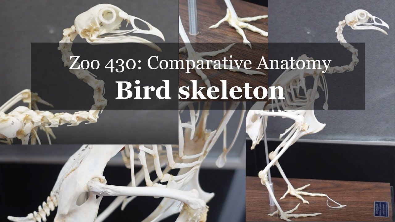
Image taken from the YouTube channel Madtown Anatomy , from the video titled Osteology: Bird skeleton walkthrough (chicken) .
Birds grace our planet with their presence, a kaleidoscope of forms and behaviors filling every corner of the Earth. From the soaring heights of eagles to the intricate dances of hummingbirds, their diversity is simply breathtaking.
But beyond the vibrant plumage and captivating songs lies a world of intricate anatomy, finely tuned by evolution to meet the demands of their varied lifestyles.
Among the many wonders of avian biology, the structure of a bird’s leg stands out as a testament to natural engineering.
Why Bird Legs Matter
Understanding the anatomy of a bird’s leg is more than just an academic exercise. It’s a gateway to appreciating the remarkable adaptations that allow these creatures to thrive in diverse environments.
Their legs are not just for walking; they are instruments of survival, perfectly shaped for perching, grasping, swimming, hunting, and so much more.
Delving into the skeletal framework reveals how these bones, muscles, and joints work in harmony to enable these behaviors.
A Guided Tour of Bird Leg Bones
This article serves as your accessible guide to the fascinating world of bird leg anatomy.
We’ll break down the key components, providing clear explanations and labeled diagrams to help you identify each bone and understand its function.
Whether you’re a seasoned ornithologist, a budding birdwatcher, or simply curious about the natural world, this journey into the avian leg promises to be both informative and engaging.
Get ready to unlock the secrets hidden within these remarkable structures, one bone at a time.
As individual marvels, each bone of the avian leg tells a story of adaptation, a narrative etched in calcium and shaped by millennia of evolutionary pressures.
But to truly grasp the genius of bird legs, we must zoom out, to see how each part functions within the bird’s overall skeletal system.
Bird Leg Anatomy: A Skeletal System Overview
The bird leg is far more than just a means of standing or walking; it’s a complex and highly specialized piece of biological machinery intricately woven into the bird’s entire skeletal structure.
Understanding this intricate relationship is key to appreciating the full scope of avian adaptation.
The Bird Skeleton: Lightweight Yet Strong
A bird’s skeleton is a masterpiece of engineering, prioritizing both strength and lightness.
Many of the bones are hollow, pneumatized with air sacs connected to the respiratory system, reducing weight for flight.
Despite this, the skeletal structure must withstand significant forces during takeoff, landing, and various terrestrial activities.
The legs, therefore, are crucial load-bearing components, directly impacted by these constraints.
The Leg’s Role in Avian Life
The leg structure is fundamental to a bird’s survival, facilitating a wide range of behaviors essential for feeding, reproduction, and predator avoidance.
Locomotion, of course, is primary.
But beyond simple walking or running, bird legs enable perching on branches, grasping prey, wading through water, and even elaborate courtship displays.
The skeletal structure of the leg directly dictates the efficiency and effectiveness of these movements.
Key Skeletal Components: A Foundation for Function
While the specific arrangement and proportions of leg bones vary widely across different avian species, the fundamental components remain consistent.
These components act as the foundation upon which specialized adaptations are built.
The femur, tibia, fibula, tarsometatarsus, and phalanges (toe bones) are the primary players in this skeletal drama.
Each bone contributes uniquely to the leg’s overall functionality.
Understanding these key anatomical features provides a solid foundation for exploring the remarkable diversity of bird leg adaptations.
The Femur: Connecting the Leg to the Body
Having established a foundational understanding of avian skeletal structure, we now turn our attention to the individual components that make up the remarkable bird leg. We’ll begin our anatomical journey at the top, with the femur.
The femur, or thigh bone, serves as the crucial link between the bird’s leg and its body. It’s more than just a static piece of bone; it’s a dynamic structure that bears weight, anchors powerful muscles, and facilitates a wide range of movements.
Location of the Femur
The femur is located in the upper part of the bird’s leg, nestled close to the body. It extends from the hip joint, where it articulates with the pelvis, to the knee joint, where it connects with the tibia and fibula.
In many bird species, the femur is relatively short and stout compared to other leg bones. This is an adaptation that contributes to a lower center of gravity, enhancing stability during flight and terrestrial activities.
Function of the Femur
The femur performs several critical functions that are vital to a bird’s survival:
-
Weight Bearing: The femur is the primary weight-bearing bone in the bird’s leg. It must withstand the forces generated during landing, walking, running, and perching. Its robust structure is designed to handle these stresses effectively.
-
Muscle Attachment: The surface of the femur serves as an attachment point for numerous powerful muscles. These muscles control a wide range of leg movements, including flexion, extension, abduction, and adduction.
-
Locomotion: By connecting the leg to the hip and knee, the femur acts as a lever that enables the bird to propel itself forward. The length and angle of the femur can influence the bird’s gait and its ability to maneuver in different environments.
Connection to the Hip Joint
The femur articulates with the pelvis at the hip joint. This is a ball-and-socket joint, which allows for a wide range of motion. The head of the femur, a rounded projection, fits into a cup-shaped socket on the pelvis called the acetabulum.
This articulation is stabilized by strong ligaments that prevent dislocation and provide support.
The hip joint plays a critical role in enabling birds to walk, run, hop, and even fly. The angle of the femur relative to the pelvis can influence a bird’s posture and its ability to balance.
The structure of the hip joint in birds is also influenced by their lifestyle. For example, birds that spend a lot of time walking or running, such as ostriches and emus, have a more robust hip joint than birds that spend most of their time flying.
Tibia and Fibula: Lower Leg Support
Having explored the crucial role of the femur in connecting the leg to the bird’s body and facilitating upper leg movement, our attention now shifts to the lower leg. Here, we encounter the tibia and fibula, two bones that work in concert to provide essential support and stability.
Location of the Tibia and Fibula
The tibia and fibula are located in the lower segment of the bird’s leg, extending from the knee joint down towards the ankle. The tibia, also known as the shinbone, is the larger and more prominent of the two. It runs parallel to the fibula, which is significantly thinner and positioned on the outer side of the leg.
Functions of the Tibia and Fibula
Tibia: Stability and Weight-Bearing
The tibia is the primary weight-bearing bone in the lower leg. Its robust structure is designed to withstand the stresses generated during locomotion and perching. The tibia also serves as a crucial attachment site for numerous muscles that control the movement of the foot and toes. These muscles are responsible for flexion, extension, and rotation of the lower leg, enabling birds to perform a wide range of activities.
Fibula: Stability and Muscle Attachment
The fibula, while smaller than the tibia, plays an important role in providing stability to the lower leg. It helps to prevent excessive lateral movement and supports the ankle joint.
The fibula also serves as an attachment point for several muscles, contributing to the overall strength and control of the lower leg. In some bird species, the fibula is reduced in size or even fused to the tibia, reflecting adaptations to specific lifestyles.
Synergistic Support
The tibia and fibula work together to provide comprehensive support for the lower leg. The tibia bears the majority of the weight, while the fibula contributes to stability and muscle attachment. Together, these two bones create a strong and flexible structure that enables birds to move efficiently on the ground, in the air, and in the water.
The arrangement of the tibia and fibula allows for a degree of independent movement, which is essential for tasks such as grasping, perching, and manipulating objects with their feet. This intricate interplay between bone structure and muscle function highlights the remarkable adaptations that have enabled birds to thrive in diverse environments.
Following the tibia and fibula, which provide structure to the lower leg, we arrive at a truly unique feature of avian anatomy – the tarsometatarsus. This specialized bone offers a fascinating glimpse into the evolutionary adaptations that have allowed birds to thrive in diverse environments.
Tarsometatarsus: A Bird-Specific Marvel
The tarsometatarsus is a bone found exclusively in birds. It is a defining characteristic of avian skeletal structure and a testament to the power of natural selection. Understanding its form and function is crucial to grasping the biomechanics of bird locomotion.
What is the Tarsometatarsus?
The tarsometatarsus is a long, slender bone located in the lower part of the bird’s leg, forming what is often mistakenly called the "ankle" or "foot." It’s important to note that the true ankle joint in birds is located higher up, between the tibia and the tarsometatarsus.
Evolutionary Origins: Fusion for Function
The tarsometatarsus isn’t just another bone; it’s a fusion of several smaller tarsal (ankle) and metatarsal (foot) bones that were present in the bird’s evolutionary ancestors. Over millions of years, these individual bones fused together to create a single, strong, and lightweight structure.
This fusion provides enhanced stability and power for activities like:
- Running
- Hopping
- Perching
- Take-off
This evolutionary process highlights how bird anatomy has been meticulously shaped to optimize flight and terrestrial movement.
Leverage and Support: The Functional Role
The primary function of the tarsometatarsus is to provide leverage and support for the foot. Acting as an extension of the lower leg, it increases the length of the limb, which is essential for efficient locomotion.
The bone’s length and robust structure allow for powerful strides during running or hopping.
It also provides a stable base for perching and grasping.
Muscles that control the toes attach to the tarsometatarsus, allowing for precise movements needed for gripping branches or capturing prey.
The tarsometatarsus is a critical component of the avian leg, enabling the diverse range of movements we observe in birds.
Adaptations in Different Bird Species
The tarsometatarsus isn’t uniform across all bird species. Its length, shape, and robustness can vary considerably, reflecting the specific lifestyle and ecological niche of a particular bird.
For example:
- Birds that spend a lot of time wading in water often have longer tarsometatarsi, which increases their leg length and allows them to navigate shallow waters more easily.
- Raptors, on the other hand, may have shorter, thicker tarsometatarsi to provide the strength and stability needed to grip and carry prey.
These variations in the tarsometatarsus demonstrate the remarkable adaptability of bird leg anatomy. It enables them to thrive in a wide array of environments.
Following the evolutionary marvel of the tarsometatarsus, the bird leg extends into a diverse array of digits, each adapted to specific ecological niches and behaviors. These digits, supported by the phalanges, showcase some of the most remarkable examples of adaptation in the avian world.
Phalanges: Toe Bones and Their Adaptations
The phalanges, or toe bones, form the distalmost part of the avian leg. Their number and arrangement are far from uniform, exhibiting incredible diversity across different bird species. These variations aren’t random; they’re finely tuned adaptations that enable birds to thrive in their respective environments.
Phalangeal Formula: A Digital Code
Each digit on a bird’s foot is composed of a varying number of phalanges. Ornithologists use a "phalangeal formula" to describe the arrangement of these bones. This formula lists the number of phalanges, starting from the innermost toe (hallux) to the outermost toe.
For example, a bird with a phalangeal formula of 2-3-4-5 would have:
- 2 phalanges on its first toe (hallux)
- 3 phalanges on its second toe
- 4 phalanges on its third toe
- 5 phalanges on its fourth toe
This formula is a valuable tool for identifying and classifying birds, as it often correlates with their lifestyle and habitat.
Variations in Toe Arrangement
While the phalangeal formula describes the number of toe bones, the arrangement of the toes themselves is equally important. Most birds have four toes, but their configuration varies significantly:
-
Anisodactyl: This is the most common arrangement, with three toes pointing forward and one pointing backward. It’s ideal for perching birds like songbirds.
-
Zygodactyl: Two toes point forward, and two point backward. This arrangement provides excellent grip for climbing, as seen in woodpeckers and parrots.
-
Heterodactyl: Similar to zygodactyl, but only found in trogons. The first two toes point backward, while the outer two point forward.
-
Syndactyl: The third and fourth toes are fused together for at least part of their length. This adaptation is common in kingfishers and some other bird groups.
-
Pamprodactyl: All four toes point forward, allowing birds to grasp cylindrical objects, such as branches. This is seen in swifts.
Adaptations for Specific Functions
The specific arrangement and structure of the phalanges directly influence a bird’s ability to perform certain tasks. Here are a few examples:
Perching
Birds with the anisodactyl arrangement are perfectly adapted for perching on branches. The backward-pointing hallux acts as a "toe lock," providing a secure grip. This allows them to rest comfortably without expending energy.
Grasping
Raptors, such as eagles and hawks, have strong, curved phalanges with sharp talons. These are essential for grasping and subduing prey. Their zygodactyl-like foot structure provides exceptional strength and stability.
Swimming
Waterfowl, like ducks and geese, have webbed feet that act as paddles. The phalanges are elongated and connected by a membrane, increasing the surface area of the foot for efficient propulsion through water. The webbing maximizes thrust and minimizes drag.
Climbing
Zygodactyl feet are perfect for climbing vertical surfaces. The two forward-pointing and two backward-pointing toes provide a secure grip on tree trunks and branches. This arrangement distributes weight evenly, reducing strain on individual toes.
Running
Some ground-dwelling birds, like ostriches, have reduced numbers of toes. This reduces the weight at the end of the leg. Also, it increases the speed and efficiency of running. Each toe is strong and robust, capable of withstanding the impact of high-speed movement.
The phalanges, seemingly simple toe bones, are a testament to the incredible diversity and adaptability of birds. Their number, arrangement, and shape reflect millions of years of evolution. This is to finely tune birds to their specific ecological niches. Studying these adaptations provides valuable insights into the relationship between form and function. Also, it increases our appreciation for the complexity of avian biology.
Following the intricate arrangement of the phalanges, and their crucial role in a bird’s interaction with its environment, it’s easy to overlook smaller, yet equally important, components of the avian leg. Among these is the patella, often understated but vital for the overall function and protection of the knee joint.
Patella (Kneecap): Protecting the Knee Joint
The patella, more commonly known as the kneecap, is a small but significant bone found in the legs of many birds. It plays a crucial role in the mechanics and protection of the knee joint. Understanding its location and function is key to appreciating the intricacies of avian locomotion.
Location of the Patella
The patella is situated at the front of the knee joint, nestled within the tendon of the quadriceps femoris muscle (though birds do not technically have a quadriceps). Its position allows it to articulate with the distal end of the femur, the thigh bone, creating a smooth gliding surface.
This strategic placement is crucial for optimizing the mechanics of leg movement.
Function: Protection and Leverage
The primary function of the patella is to protect the knee joint from injury. By acting as a shield, it disperses forces that would otherwise concentrate on the joint capsule and ligaments. This is particularly important in birds, whose legs undergo significant stress during activities like landing, perching, and running.
Beyond protection, the patella also improves the efficiency of the extensor muscles of the leg.
It acts as a fulcrum, increasing the angle at which the tendon pulls on the tibia.
This, in turn, enhances the leverage of the muscles, allowing the bird to generate more force with less effort. The patella essentially optimizes the biomechanics of the knee joint, facilitating powerful and efficient movements.
Patellar Variations
While the function of the patella remains consistent across bird species, its size and shape can vary depending on the bird’s lifestyle and locomotive demands. Birds that engage in more demanding activities, such as soaring birds or strong runners, may have a relatively larger and more robust patella to withstand greater forces. Conversely, smaller birds or those with less strenuous lifestyles may have a proportionally smaller patella.
Ultimately, the patella, though small, plays a significant role in maintaining the health and functionality of the avian leg.
Following the intricate arrangement of the phalanges, and their crucial role in a bird’s interaction with its environment, it’s easy to overlook smaller, yet equally important, components of the avian leg. Among these is the patella, often understated but vital for the overall function and protection of the knee joint.
Muscles and Joints: The Mechanics of Bird Leg Movement
The skeletal framework of the bird leg, while providing structure and support, is inherently static. It is the interplay of muscles and joints that breathes life into this framework, transforming it into a dynamic system capable of a remarkable range of movements. Understanding this synergy is crucial to appreciating the biomechanical sophistication of avian locomotion.
The Musculoskeletal Partnership
Muscles act as the engines of movement, contracting to exert force on bones, while joints serve as the hinges, allowing bones to pivot and rotate relative to one another. This partnership enables birds to walk, hop, perch, fly (in some cases taking off from the ground), and perform other specialized movements.
The architecture of each joint, coupled with the specific arrangement and power of the surrounding muscles, dictates the types of motion possible.
Key Muscle Groups and Their Actions
Several key muscle groups are responsible for the primary movements of the bird leg: flexion, extension, and rotation.
Flexion
Flexion refers to the bending of a joint, decreasing the angle between two bones. In the bird leg, flexion is primarily driven by muscles located on the cranial (front) aspect of the leg. These muscles, upon contraction, pull the lower leg towards the thigh, enabling movements like crouching or bringing the foot up towards the body during flight.
Extension
Extension is the opposite of flexion, involving the straightening of a joint and increasing the angle between bones. Extension of the bird leg is primarily achieved by muscles located on the caudal (rear) aspect of the leg. These muscles are crucial for powerful movements like pushing off the ground during takeoff or extending the leg for landing.
Rotation
Rotation involves the twisting of a bone around its long axis. While less pronounced in the bird leg compared to flexion and extension, rotation is still important for fine-tuning movements and maintaining balance. Muscles responsible for rotation are strategically positioned around the hip and knee joints, allowing for subtle adjustments in leg position.
The Role of Tendons and Ligaments
It’s important to acknowledge the supporting roles of tendons and ligaments in this system.
Tendons, composed of strong, fibrous connective tissue, connect muscles to bones, transmitting the force generated by muscle contractions.
Ligaments, similarly strong connective tissues, connect bone to bone, providing stability to joints and preventing excessive or unnatural movements. These structures work in concert with the muscles and joints to ensure smooth, coordinated, and controlled leg movements.
Following the intricate arrangement of the phalanges, and their crucial role in a bird’s interaction with its environment, it’s easy to overlook smaller, yet equally important, components of the avian leg. Among these is the patella, often understated but vital for the overall function and protection of the knee joint.
With the complex interplay of musculature and skeletal structure in mind, one might reasonably ask: How does this leg structure differ across the vast spectrum of avian species?
Comparative Anatomy: Bird Leg Variations Across Species
The avian world is a testament to evolutionary adaptation, and nowhere is this more evident than in the diverse forms of bird legs. From the talons of an eagle to the webbed feet of a duck, the skeletal structure of a bird’s leg is exquisitely tailored to its specific lifestyle and ecological niche.
This section explores these variations, highlighting how bone length, shape, and overall leg architecture reflect the unique demands placed upon different avian species.
Raptors: The Power of Predation
Raptors, such as eagles, hawks, and owls, are masters of aerial hunting. Their legs are a study in power and precision. The tarsometatarsus is typically short and thick, providing exceptional strength for gripping prey.
The phalanges are equipped with sharp, curved talons, capable of delivering a lethal blow and securing even the most elusive quarry. The bone structure is dense and robust, built to withstand the forces generated during high-speed dives and struggles with prey.
These adaptations are not merely cosmetic; they are fundamental to the raptor’s predatory success.
Waterfowl: Navigating the Aquatic Realm
In stark contrast to the raptors, waterfowl like ducks, geese, and swans have evolved legs optimized for aquatic locomotion. Their tibias and fibulas are often proportionally longer, contributing to powerful paddling strokes.
The most distinctive feature, however, is the presence of webbing between the phalanges. This webbing increases the surface area of the foot, transforming it into an efficient paddle for propelling the bird through water.
Additionally, the legs are positioned further back on the body, enhancing stability and maneuverability in an aquatic environment.
Perching Birds: Masters of Balance
Perching birds, including songbirds, finches, and robins, exhibit a leg structure designed for grasping branches and maintaining balance.
The hallux (the equivalent of a big toe) is typically positioned at the back of the foot, allowing the bird to grip a perch securely. This anisodactyl foot arrangement is a defining characteristic of perching birds.
Tendons within the leg automatically tighten when the bird lands on a branch, providing a secure grip without requiring constant muscular effort. This specialized adaptation allows perching birds to rest comfortably for extended periods, even while sleeping.
Other Notable Adaptations
Beyond these examples, a myriad of other avian leg adaptations exist.
- Wading birds, like herons and egrets, possess elongated legs for navigating shallow waters.
- Ground-dwelling birds, such as ostriches and emus, have powerful legs built for running at high speeds.
- Climbing birds, such as woodpeckers, feature zygodactyl feet (two toes pointing forward and two pointing backward) for gripping vertical surfaces.
Bone Length and Shape: A Reflection of Lifestyle
Ultimately, the variations in bird leg structure underscore the principle of adaptation. Bone length, shape, and the arrangement of phalanges are all subject to selective pressures, resulting in a diverse array of forms perfectly suited to the demands of each species’ unique lifestyle. By studying these adaptations, we gain a deeper appreciation for the evolutionary forces that have shaped the avian world.
Why Bird Leg Anatomy Matters: Ornithology and Veterinary Applications
The study of bird leg anatomy extends far beyond academic curiosity. A thorough comprehension of avian skeletal structure, specifically within the leg, proves invaluable across diverse fields, from advancing ornithological research to enhancing veterinary practices. Understanding how these intricate systems function is not just about knowing the names of bones; it’s about unlocking insights into avian evolution, behavior, and health.
Ornithology and Avian Research: A Foundation for Discovery
In ornithology, a robust understanding of bird leg anatomy serves as a foundational tool for various research endeavors. Comparative studies of leg structures across different avian species offer valuable insights into evolutionary relationships and adaptive strategies.
For instance, the length and robustness of the tarsometatarsus can reveal crucial information about a bird’s locomotion style, habitat preference, and even foraging behavior. Analyzing these skeletal features contributes significantly to our understanding of avian phylogeny and ecological adaptation.
Furthermore, leg morphology can be used to study the impact of environmental changes on bird populations. Researchers can assess the effects of pollution, habitat loss, or climate change by examining skeletal abnormalities or variations in bone density. These analyses often serve as vital indicators of broader ecosystem health.
Veterinary Medicine: Diagnosis, Treatment, and Rehabilitation
For veterinarians specializing in avian care, a comprehensive knowledge of bird leg anatomy is paramount for accurate diagnosis, treatment, and rehabilitation of injuries and diseases.
Avian legs are susceptible to a variety of conditions, including fractures, dislocations, tendon ruptures, and arthritis. A precise understanding of the underlying skeletal and muscular structure is essential for effective diagnosis using radiographs, palpation, and other diagnostic techniques.
Moreover, specialized knowledge of avian bone structure is crucial for performing surgical procedures, such as fracture repair or joint stabilization. The delicate nature of bird bones requires careful handling and precise techniques to ensure successful outcomes. Post-operative care and rehabilitation also heavily rely on an understanding of the leg’s biomechanics to restore proper function and mobility.
Enhancing Avian Welfare Through Anatomical Knowledge
By leveraging our knowledge of bird leg anatomy, veterinary professionals can significantly enhance the welfare of avian patients, both in clinical settings and in conservation efforts.
Appreciation for Avian Biology: Form, Function, and Beauty
Beyond its practical applications, understanding bird leg anatomy fosters a deeper appreciation for the remarkable adaptations and evolutionary marvels present in the avian world. The elegant design of bird legs, each bone perfectly shaped and positioned to fulfill its specific role, reflects the power of natural selection in sculpting form to optimize function.
From the powerful talons of an eagle to the delicate feet of a songbird, each avian leg tells a story of adaptation and survival. Studying these intricate structures allows us to connect with the natural world on a deeper level, recognizing the inherent beauty and complexity of avian biology. This deeper understanding, in turn, fosters a greater commitment to conservation and stewardship of our planet’s avian diversity.
Labeled Bird Leg Bone Diagrams: A Visual Guide
Having explored the individual components of a bird’s leg, from the femur to the phalanges, it’s time to consolidate that knowledge.
Visual aids are invaluable tools for anatomical understanding, especially when dealing with complex structures like the avian leg.
This section offers detailed, labeled diagrams and illustrations designed to clarify the location and relationships of each bone.
High-Quality Visualizations for Effective Learning
The efficacy of anatomical study hinges on the quality of the visuals used.
Poorly rendered or inaccurately labeled diagrams can lead to confusion and hinder comprehension.
Our diagrams are meticulously crafted to provide clear, detailed representations of the avian leg skeleton.
We prioritize anatomical accuracy, ensuring that each bone is depicted in its correct shape, proportion, and orientation.
Furthermore, the illustrations are designed to be visually appealing and easy to interpret, making the learning process more engaging and less daunting.
Key Bones Clearly Labeled
The following bones are prominently featured and clearly labeled in our diagrams:
- Femur: The thigh bone, connecting the leg to the body.
- Tibia: The main bone of the lower leg.
- Fibula: A slender bone alongside the tibia, providing additional support.
- Tarsometatarsus: The unique, fused bone specific to birds, forming part of the lower leg and foot.
- Phalanges: The toe bones, adapted for various functions.
These labels are strategically placed to avoid cluttering the image while remaining easily visible and directly associated with the corresponding bone.
Understanding Anatomical Relationships
Beyond simply identifying individual bones, the diagrams also highlight their spatial relationships.
Understanding how these bones connect and interact is crucial for grasping the biomechanics of the bird leg.
The illustrations demonstrate the articulations at the hip, knee, and ankle joints, providing context for understanding how these bones move together.
Different Views for Comprehensive Understanding
To provide a complete understanding of bird leg anatomy, we include multiple views in our diagrams.
These may include:
- Lateral (side) view
- Anterior (front) view
- Posterior (back) view
These various perspectives allow you to visualize the three-dimensional structure of the leg and appreciate the subtle curvatures and features of each bone.
The inclusion of different viewpoints solidifies the overall understanding of bird leg bone structures.
Bird Leg Bones Labeled: Frequently Asked Questions
Here are some common questions we receive about bird leg anatomy and understanding the bird leg bones labeled in our guide.
What’s the difference between the ‘tibia’ and ‘fibula’ in a bird’s leg?
In bird leg bones labled, the tibia (or tibiotarsus, actually) is the larger, main bone in the lower leg, akin to our shinbone. The fibula is much smaller and thinner, running alongside the tibia. In many adult birds, it’s significantly reduced in size.
Why is the "ankle" bone labeled as the tarsometatarsus?
Birds don’t have a distinct ankle joint like humans. Instead, the bones of the ankle and foot have fused into a single bone called the tarsometatarsus. When bird leg bones labeled illustrations show the "ankle," it’s actually this fused structure.
What is the function of the patella (kneecap) in a bird’s leg?
The patella, or kneecap, in a bird’s leg functions similarly to ours. It protects the knee joint and improves the leverage of the thigh muscles. While sometimes less prominent, the bird leg bones labeled illustrations highlight its presence.
Where does the femur fit into the bird’s body?
The femur is the bird’s thigh bone, connecting the hip joint to the knee. It’s often hidden by feathers. In bird leg bones labeled diagrams, it shows the starting point of the leg structure within the bird’s overall anatomy.
So, that’s your quick guide to birdleg bones labled! Hopefully, you now have a better understanding of how these amazing structures work. Now get out there and see if you can spot these bones in action!
