Bacteria Cell Labeled: Unlock the Microscopic World!
The structure of a bacteria cell labeled reveals critical insights into its function. Microscopy, a fundamental technique, enables scientists to visualize the intricate components within bacteria. Understanding these components is crucial for researchers at institutions like the Centers for Disease Control and Prevention (CDC), as it aids in developing effective treatments for bacterial infections. Cell walls, key structures often seen on any bacteria cell labeled, provide structural support and protection to bacterial cells.
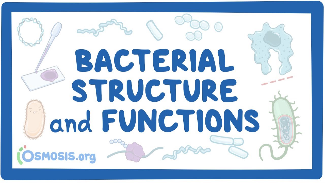
Image taken from the YouTube channel Osmosis from Elsevier , from the video titled Bacterial Structure and Functions .
Bacteria are arguably the most ubiquitous and diverse forms of life on Earth. They inhabit virtually every environment, from the deepest ocean trenches to the driest deserts, playing crucial roles in nutrient cycling, decomposition, and even influencing global climate patterns.
Their significance extends far beyond natural ecosystems, impacting fields like medicine, agriculture, and biotechnology. Understanding these microscopic organisms is paramount to addressing some of humanity’s most pressing challenges.
The Power of Labeling: Illuminating the Invisible
Gaining insights into the intricacies of bacterial cells presents a significant challenge due to their microscopic size and complex internal organization. This is where the power of labeling techniques comes into play.
Labeling bacterial cells involves using specific markers, often fluorescent dyes or antibodies, to tag and visualize particular cellular components. These markers bind to specific structures, allowing scientists to observe their location, interactions, and dynamics within the cell.
This process allows us to observe cellular processes in action.
The impact of labeling techniques on our understanding of bacterial cells cannot be overstated. It has revolutionized our ability to study bacterial cell structure and function.
It also allows us to characterize different bacterial species.
By visualizing these differences, we can better understand bacterial physiology, genetics, and interactions with their environment.
Thesis Statement: A Journey into the Labeled Bacterial Cell
This exploration of the bacterial world through labeling techniques is more than just a visual exercise. It’s an avenue to deeper knowledge.
A comprehensive exploration of the labeled bacterial cell reveals key components and differences, providing insights into their function and classification.
Through this lens, we can understand the core components of bacterial life and the subtle variations that enable bacteria to thrive in diverse conditions. These understandings will help refine classification methods of bacterial life.
Gaining insights into the intricacies of bacterial cells presents a significant challenge due to their microscopic size and complex internal organization. This is where the power of labeling techniques comes into play.
Labeling bacterial cells involves using specific markers, often fluorescent dyes or antibodies, to tag and visualize particular cellular components. These markers bind to specific structures, allowing scientists to observe their location, interactions, and dynamics within the cell.
This process allows us to observe cellular processes in action.
The impact of labeling techniques on our understanding of bacterial cells cannot be overstated. It has revolutionized our ability to study bacterial cell structure and function.
It also allows us to characterize different bacterial species.
By visualizing these differences, we can better understand bacterial physiology, genetics, and interactions with their environment.
Thus equipped with the tools to see the unseen, we turn our attention to one of the most critical structures defining bacterial life: the cell wall.
The Bacterial Fortress: Exploring the Cell Wall
The bacterial cell wall is an essential and dynamic structure that dictates cell shape, offers robust protection, and maintains structural integrity. It is a complex architecture that shields the delicate inner workings of the bacterial cell from a hostile external environment.
This fortress-like barrier is not merely a passive shield. It actively participates in interactions with the surrounding environment, influencing bacterial survival and pathogenicity.
The Rigid Framework: Shape and Protection
The cell wall’s most fundamental function is providing a rigid framework that defines the bacterium’s characteristic shape. Whether it’s the spherical coccus, the rod-shaped bacillus, or the spiral spirillum, the cell wall dictates its morphology.
Beyond shape, the cell wall serves as a crucial protective barrier. It safeguards the cell against osmotic pressure, preventing it from bursting in hypotonic environments.
It also provides a defense against mechanical stress and the intrusion of harmful substances.
The Keystone: Peptidoglycan’s Crucial Role
At the heart of the bacterial cell wall lies peptidoglycan, a unique and complex polymer also known as murein. This mesh-like structure is found exclusively in bacteria and is essential for their survival.
Peptidoglycan is composed of glycan chains, alternating N-acetylglucosamine (NAG) and N-acetylmuramic acid (NAM), cross-linked by short peptides. This intricate cross-linking provides the cell wall with its remarkable strength and rigidity.
The precise composition and structure of peptidoglycan can vary between bacterial species, influencing their susceptibility to antibiotics and other antimicrobial agents.
Gram-Positive vs. Gram-Negative: A Tale of Two Walls
One of the most fundamental distinctions in bacteriology is the division of bacteria into two major groups: Gram-positive and Gram-negative. This classification is based on differences in their cell wall structure, revealed through the Gram staining procedure.
The contrasting cell wall architectures of these two groups have profound implications for their physiology, pathogenicity, and antibiotic resistance.
Gram-Positive Bacteria: A Thick Peptidoglycan Layer
Gram-positive bacteria are characterized by a thick peptidoglycan layer, ranging from 20 to 80 nanometers in thickness. This thick layer forms the outermost layer of the cell wall, providing a robust barrier against the external environment.
Embedded within this peptidoglycan matrix are teichoic acids, unique polymers that contribute to the cell wall’s rigidity and play a role in cell adhesion and biofilm formation.
Teichoic acids can also act as antigens, triggering immune responses in host organisms.
Gram-Negative Bacteria: A Thin Layer and Outer Membrane
In contrast to Gram-positive bacteria, Gram-negative bacteria possess a thin peptidoglycan layer, typically only 5 to 10 nanometers thick.
This peptidoglycan layer is located in the periplasmic space, a gel-like region between the inner cell membrane and the outer membrane.
The defining feature of Gram-negative bacteria is the presence of an outer membrane, a unique lipid bilayer composed of phospholipids, proteins, and lipopolysaccharide (LPS).
LPS, also known as endotoxin, is a potent immunostimulant that can trigger strong inflammatory responses in animals. It is a major virulence factor in many Gram-negative pathogens.
The outer membrane also provides an additional barrier against antibiotics and other antimicrobial agents, contributing to the increased antibiotic resistance often observed in Gram-negative bacteria.
Understanding the structural differences between Gram-positive and Gram-negative cell walls is crucial for developing effective strategies to combat bacterial infections. The unique features of each cell wall type represent potential targets for novel antimicrobial agents.
The cell wall, with its robust architecture, provides the first line of defense and establishes the bacterium’s identity. But beneath this protective layer lies the cell membrane and the cytoplasm, equally vital components that orchestrate essential life processes.
These structures are the gatekeepers and powerhouses of the bacterial cell, responsible for regulating the flow of nutrients, generating energy, and housing the genetic material that dictates the bacterium’s destiny.
Gatekeeper and Powerhouse: The Cell Membrane and Cytoplasm
The bacterial cell membrane, also known as the plasma membrane, is a selectively permeable barrier that surrounds the cytoplasm. It’s a dynamic structure that regulates the movement of substances in and out of the cell, ensuring the proper internal environment for cellular processes.
The Cell Membrane: A Selective Barrier
The cell membrane is primarily composed of a phospholipid bilayer, similar to eukaryotic cell membranes. This bilayer consists of two layers of phospholipid molecules, each with a hydrophilic (water-attracting) head and a hydrophobic (water-repelling) tail.
This arrangement creates a barrier that is impermeable to most water-soluble molecules, while allowing the passage of small, nonpolar molecules.
Embedded within the phospholipid bilayer are various proteins that perform a multitude of functions, including transport, cell signaling, and energy production.
Regulating Transport: The cell membrane regulates the transport of nutrients, ions, and waste products across the membrane.
This is achieved through various mechanisms, including:
-
Passive transport: This process does not require energy and relies on the concentration gradient to move substances across the membrane. Examples include diffusion and osmosis.
-
Active transport: This process requires energy, usually in the form of ATP, to move substances against their concentration gradient. This is often mediated by specific transport proteins.
-
Group translocation: This unique transport mechanism involves the chemical modification of the transported substance as it crosses the membrane.
Energy Production and Cell Signaling
Beyond its role in transport, the cell membrane plays a crucial role in energy production. In bacteria, the cell membrane houses the electron transport chain, a series of protein complexes that generate a proton gradient across the membrane.
This proton gradient is then used to drive the synthesis of ATP, the cell’s primary energy currency, through a process called oxidative phosphorylation.
The cell membrane also participates in cell signaling, allowing bacteria to sense and respond to changes in their environment.
Receptor proteins on the cell membrane bind to specific signaling molecules, triggering a cascade of intracellular events that ultimately alter gene expression or cellular behavior.
The Cytoplasm: The Cell’s Interior
The cytoplasm is the gel-like substance that fills the interior of the bacterial cell. It is composed primarily of water, but also contains a variety of other molecules, including:
-
Proteins: Enzymes, structural proteins, and transport proteins.
-
Carbohydrates: Energy sources and building blocks.
-
Lipids: Membrane components and energy storage molecules.
-
Inorganic ions: Essential for various cellular processes.
The cytoplasm is the site of many essential metabolic reactions, including glycolysis, the citric acid cycle, and protein synthesis. It also houses the bacterial chromosome and ribosomes.
The Nucleoid Region: Organizing Bacterial DNA
Unlike eukaryotic cells, bacteria lack a membrane-bound nucleus. Instead, their DNA is organized within a region of the cytoplasm called the nucleoid.
The nucleoid is not a defined structure, but rather a densely packed area containing the bacterial chromosome. The bacterial chromosome is typically a single, circular DNA molecule that contains all the genetic information necessary for the cell’s survival and reproduction.
The DNA within the nucleoid is tightly packed and organized with the help of various proteins, including nucleoid-associated proteins (NAPs). This compact structure allows the long DNA molecule to fit within the confines of the bacterial cell.
Protein Factories: Unveiling the Role of Ribosomes
With a grasp on the cell membrane’s selective nature and the cytoplasm’s bustling environment, we now turn our attention to the intricate machinery responsible for building the very proteins that drive bacterial life: the ribosomes.
The Architecture of Bacterial Ribosomes
Bacterial ribosomes, the workhorses of protein synthesis, are complex molecular machines found within the cytoplasm. These structures are responsible for translating genetic code into functional proteins, crucial for bacterial survival and function.
Unlike their eukaryotic counterparts, bacterial ribosomes are characterized by their 70S sedimentation coefficient, consisting of two subunits: a smaller 30S subunit and a larger 50S subunit.
The 30S subunit is composed of a single ribosomal RNA (rRNA) molecule and approximately 21 ribosomal proteins.
The 50S subunit comprises two rRNA molecules and about 34 ribosomal proteins.
These subunits assemble around messenger RNA (mRNA) during translation, forming a functional ribosome capable of synthesizing proteins.
Decoding the Blueprint: The Process of Translation
Translation, the process of protein synthesis, is a highly coordinated event that occurs on the ribosome. It involves the decoding of mRNA to assemble a specific sequence of amino acids into a polypeptide chain.
The process begins with the binding of the 30S subunit to the mRNA, followed by the recruitment of the initiator tRNA carrying the first amino acid, typically formylmethionine (fMet).
The 50S subunit then joins the complex, forming the complete 70S ribosome.
As the ribosome moves along the mRNA, each codon (a sequence of three nucleotides) is recognized by a specific transfer RNA (tRNA) molecule carrying the corresponding amino acid.
The ribosome catalyzes the formation of peptide bonds between amino acids, elongating the polypeptide chain.
Finally, when the ribosome encounters a stop codon on the mRNA, translation terminates, releasing the completed polypeptide chain.
Ribosomes: Cornerstones of Bacterial Life
The significance of ribosomes extends far beyond their role in protein synthesis. They are essential for bacterial growth, metabolism, and survival.
Proteins synthesized by ribosomes are involved in virtually every aspect of bacterial life, including:
- Enzymatic reactions: Catalyzing biochemical processes.
- Structural support: Providing shape and stability to the cell.
- Transport: Facilitating the movement of molecules across the cell membrane.
- Regulation: Controlling gene expression and cellular processes.
Furthermore, ribosomes are targets for many antibiotics.
Drugs like tetracycline and erythromycin inhibit bacterial protein synthesis by binding to the ribosome and disrupting its function. This makes ribosomes a critical target for combating bacterial infections.
The efficient and accurate synthesis of proteins by ribosomes is crucial for maintaining cellular homeostasis and adapting to changing environmental conditions, underscoring their importance in bacterial survival.
With the inner workings of the bacterial cell – from protein synthesis at the ribosomes to the organization of DNA in the nucleoid – understood, it’s time to venture beyond the cell’s surface. We now turn our attention to the various external structures that enable bacteria to interact with their environment, move, adhere to surfaces, and even resist harsh conditions.
Appendages and Armor: External Structures of Bacteria
Bacteria are not simply passive entities floating in a nutrient broth. They are active participants in their environments, equipped with a variety of external structures that facilitate motility, attachment, and protection. These appendages and protective layers play critical roles in bacterial survival, pathogenicity, and the formation of complex communities known as biofilms.
Flagella: Propelling Bacterial Movement
Flagella are whip-like appendages that enable bacteria to move through their environment.
These structures are not simply passive propellers; they are complex molecular motors that rotate to generate thrust.
The basic structure of a bacterial flagellum consists of three main components:
-
The filament, a long, helical structure composed of the protein flagellin.
-
The hook, a flexible connector between the filament and the basal body.
-
The basal body, a complex motor embedded in the cell membrane and cell wall.
Types of Flagellar Arrangements
The arrangement of flagella on a bacterial cell can vary, leading to different types of motility.
-
Monotrichous: A single flagellum at one pole of the cell.
-
Amphitrichous: A single flagellum at both poles of the cell.
-
Lophotrichous: A tuft of flagella at one or both poles of the cell.
-
Peritrichous: Flagella distributed over the entire surface of the cell.
These arrangements influence the bacterium’s swimming behavior, affecting its ability to navigate towards nutrients or away from harmful substances.
Pili (Fimbriae): Attachment and Biofilm Formation
Pili, also known as fimbriae, are short, hair-like appendages that extend from the bacterial cell surface.
Unlike flagella, pili are primarily involved in attachment to surfaces, including host cells and other bacteria.
These structures are composed of protein subunits called pilins, which assemble to form a thin, filamentous structure.
The Role of Pili in Biofilm Formation and Pathogenicity
Pili play a crucial role in the formation of biofilms, which are structured communities of bacteria encased in a self-produced matrix.
By adhering to surfaces and to each other, bacteria can form biofilms that are more resistant to antibiotics and immune responses.
In pathogenic bacteria, pili can also mediate attachment to host cells, a critical step in the infection process.
Specific types of pili, such as type IV pili, can even facilitate motility through a mechanism called twitching motility.
The Capsule: A Protective Shield
The capsule is a layer of polysaccharide or protein that surrounds the bacterial cell wall.
This structure provides a protective barrier against various environmental stresses, including desiccation and phagocytosis by immune cells.
The capsule is often described as a "slime layer" because of its hydrated and gelatinous nature.
Capsule’s Role in Virulence and Survival
The capsule enhances bacterial virulence by interfering with the host’s immune system.
By preventing phagocytosis, the capsule allows bacteria to evade destruction by immune cells and establish infection.
Additionally, the capsule can contribute to biofilm formation by promoting adhesion and providing a structural framework for the community.
Endospores: Survival Under Stress
Unlike the other structures discussed, endospores are not external appendages but rather dormant forms of bacteria that are highly resistant to harsh conditions.
Endospores are formed in response to environmental stress, such as nutrient deprivation or exposure to extreme temperatures or radiation.
Formation and Structure of Endospores
The process of endospore formation, or sporulation, involves a complex series of morphological and biochemical changes within the bacterial cell.
The resulting endospore contains a copy of the bacterial chromosome, essential proteins, and a high concentration of dipicolinic acid, a compound that contributes to heat resistance.
The endospore is encased in a tough outer coat that provides protection against various environmental stressors.
When conditions become favorable, the endospore can germinate, giving rise to a new vegetative bacterial cell.
With the inner workings of the bacterial cell – from protein synthesis at the ribosomes to the organization of DNA in the nucleoid – understood, it’s time to venture beyond the cell’s surface. We now turn our attention to the various external structures that enable bacteria to interact with their environment, move, adhere to surfaces, and even resist harsh conditions.
Gram-Positive vs. Gram-Negative: A Tale of Two Walls
The bacterial world is broadly divided into two major groups based on a fundamental difference: the structure of their cell walls. This distinction, revealed by the Gram stain, separates bacteria into Gram-positive and Gram-negative categories, each possessing unique characteristics that impact their susceptibility to antibiotics, their interactions with host organisms, and their overall ecological roles. Understanding these differences is crucial for fields ranging from medicine to environmental science.
Defining Gram-Positive Bacteria
Gram-positive bacteria are characterized by a thick peptidoglycan layer that constitutes a significant portion of their cell wall. This robust layer, composed of cross-linked chains of sugars and amino acids, provides structural integrity and protection against osmotic pressure.
Embedded within this peptidoglycan matrix are teichoic acids, unique polymers that extend outward from the cell wall. Teichoic acids play several crucial roles, including:
- Regulating cell wall turnover.
- Participating in cell division.
- Potentially contributing to adhesion to surfaces.
- Eliciting an immune response in host organisms.
The cell wall of Gram-positive bacteria is relatively simple, lacking an outer membrane. This feature makes them generally more susceptible to certain antibiotics, such as penicillin, that target peptidoglycan synthesis.
Unveiling Gram-Negative Bacteria
In contrast to their Gram-positive counterparts, Gram-negative bacteria possess a more complex cell wall structure. They have a thin peptidoglycan layer, which is located in the periplasmic space between the inner cell membrane and the outer membrane.
The outer membrane is a defining feature of Gram-negative bacteria. This membrane contains lipopolysaccharide (LPS), a potent endotoxin that can trigger a strong immune response in animals, leading to inflammation and even septic shock. LPS is composed of three parts:
- Lipid A: The hydrophobic anchor embedded in the outer membrane, responsible for the toxic effects of LPS.
- Core oligosaccharide: A short chain of sugars linked to Lipid A.
- O-antigen: A highly variable polysaccharide chain that extends outward from the cell surface, contributing to serotype diversity and acting as an antigen.
The periplasmic space is a gel-like compartment located between the inner and outer membranes of Gram-negative bacteria. It contains a variety of proteins involved in nutrient transport, protein folding, and degradation of toxins.
The outer membrane of Gram-negative bacteria acts as a permeability barrier, making them generally more resistant to antibiotics and detergents than Gram-positive bacteria.
Comparative Analysis: A Structural Showdown
The key difference between Gram-positive and Gram-negative bacteria lies in the structure and composition of their cell walls. The thick peptidoglycan layer of Gram-positive bacteria provides strength and rigidity, while the outer membrane of Gram-negative bacteria offers protection against environmental stressors and antimicrobial agents.
| Feature | Gram-Positive Bacteria | Gram-Negative Bacteria |
|---|---|---|
| Peptidoglycan Layer | Thick | Thin |
| Outer Membrane | Absent | Present, containing Lipopolysaccharide (LPS) |
| Teichoic Acids | Present | Absent |
| Periplasmic Space | Narrow | Wide |
| Antibiotic Resistance | Generally more susceptible | Generally more resistant |
| Gram Stain | Retains crystal violet (purple/blue) | Loses crystal violet, counterstained by safranin (pink/red) |
Understanding these structural differences is not merely an academic exercise. It has profound implications for:
- Medical diagnostics: The Gram stain is a rapid and inexpensive method for identifying bacteria and guiding antibiotic therapy.
- Drug development: The unique cell wall structures of Gram-positive and Gram-negative bacteria are targets for new antimicrobial agents.
- Infection control: Understanding the mechanisms of antibiotic resistance in different bacterial groups is crucial for preventing the spread of infections.
- Biotechnology: Bacterial cell walls can be engineered for various applications, such as drug delivery and vaccine development.
In conclusion, the tale of two walls – Gram-positive and Gram-negative – reveals a fundamental dichotomy in the bacterial world. These structural differences not only define the classification of bacteria but also dictate their interactions with the environment and their impact on human health. By unraveling the intricacies of bacterial cell wall structure, we gain valuable insights that can be harnessed to combat infectious diseases and develop innovative biotechnological solutions.
Seeing is Believing: Microscopy and Staining Techniques
Understanding the intricate world of bacteria hinges on our ability to visualize these microscopic entities. While the knowledge of cellular components is vital, translating that knowledge into tangible observations requires the application of various microscopy and staining techniques. These techniques serve as indispensable tools, bridging the gap between theoretical understanding and empirical observation, allowing us to identify, classify, and study bacteria in unprecedented detail.
A Window into the Microscopic World: Microscopy Techniques
Microscopy serves as the fundamental method for directly observing bacteria, overcoming the limitations of the naked eye. Different types of microscopy offer unique capabilities, allowing researchers to investigate bacterial structures at various levels of magnification and resolution.
Light Microscopy: The Foundation of Bacterial Observation
Light microscopy, including brightfield, darkfield, and phase contrast microscopy, represents the cornerstone of bacterial visualization. Brightfield microscopy, the most common technique, illuminates the sample from below, allowing us to observe stained or naturally pigmented bacteria.
Darkfield microscopy enhances contrast by illuminating the sample from the sides, making unstained bacteria appear bright against a dark background. Phase contrast microscopy exploits differences in refractive index within the cell to create contrast, revealing internal structures without the need for staining.
Electron Microscopy: Unveiling the Ultrastructure
Electron microscopy takes visualization to the nanometer scale. Transmission electron microscopy (TEM) transmits a beam of electrons through a thinly prepared sample, revealing the internal ultrastructure of bacterial cells with exceptional detail.
Scanning electron microscopy (SEM), on the other hand, scans the surface of the sample with a focused electron beam, generating high-resolution images of bacterial morphology and surface features.
Fluorescence Microscopy: Illuminating Specific Structures
Fluorescence microscopy uses fluorescent dyes or proteins (fluorophores) to selectively label specific bacterial cell structures. When exposed to specific wavelengths of light, these fluorophores emit light of a longer wavelength, enabling researchers to visualize the location and distribution of target molecules within the cell. This method is invaluable for studying dynamic processes and interactions within bacterial cells.
Painting a Cellular Portrait: Fluorescent Labeling
The power of fluorescence microscopy lies in its ability to target and illuminate specific components within a bacterial cell. Fluorescent dyes can be conjugated to antibodies that recognize specific surface proteins or internal molecules, enabling researchers to selectively label these targets.
Genetic engineering techniques allow for the creation of bacteria that express fluorescent proteins, such as green fluorescent protein (GFP), fused to specific proteins of interest. This allows for the real-time visualization of protein localization and dynamics within living cells.
The Art of Staining: Differentiating and Identifying Bacteria
While microscopy provides the means to visualize bacteria, staining techniques enhance contrast and enable the differentiation of bacterial species based on their structural and chemical properties.
Gram Staining: A Cornerstone of Bacterial Classification
The Gram stain, developed by Hans Christian Gram, remains one of the most widely used and fundamental staining techniques in microbiology. This differential staining method categorizes bacteria into two major groups: Gram-positive and Gram-negative, based on differences in their cell wall structure.
Gram-positive bacteria retain the crystal violet stain due to their thick peptidoglycan layer, appearing purple under the microscope. Gram-negative bacteria, with their thinner peptidoglycan layer and outer membrane, lose the crystal violet stain during decolorization and are counterstained with safranin, appearing pink. This simple yet powerful staining procedure provides crucial information for bacterial identification and diagnosis.
Beyond Gram Staining: Highlighting Specific Structures
In addition to Gram staining, other staining methods are employed to visualize specific bacterial structures that may not be readily apparent with general staining techniques.
Endospore staining, for example, utilizes heat to force dye into the resistant endospores produced by certain bacteria, differentiating spore-forming bacteria from vegetative cells. Capsule staining employs a combination of acidic and basic dyes to visualize the capsule, a protective layer surrounding some bacteria. These specialized staining methods provide valuable insights into the unique characteristics of different bacterial species.
Electron microscopy and staining techniques bring the unseen world of bacteria into sharp focus, enabling detailed observations and classifications. But how does this knowledge translate into practical applications? The study of model organisms provides invaluable insights into bacterial behavior, genetics, and pathogenicity. By examining well-characterized species, researchers can unravel the complexities of bacterial life and develop strategies to combat harmful infections.
Model Organisms: Case Studies of E. coli and S. aureus
Escherichia coli (E. coli) and Staphylococcus aureus are two of the most extensively studied bacteria, serving as vital models for understanding fundamental biological processes and addressing critical healthcare challenges. Their contrasting characteristics and roles highlight the diversity within the bacterial world and the importance of in-depth research on specific species.
Escherichia coli: The Workhorse of Molecular Biology
E. coli is a Gram-negative bacterium commonly found in the lower intestine of warm-blooded organisms. While some strains are pathogenic, causing food poisoning and other illnesses, the vast majority are harmless commensals. E. coli‘s significance stems from its ease of cultivation, rapid growth rate, and well-characterized genetics.
A Cornerstone of Genetic Research
E. coli has been instrumental in advancing our understanding of molecular biology and genetics. Its relatively simple genome and efficient replication mechanisms make it an ideal model for studying DNA replication, gene expression, and protein synthesis. The bacterium has been widely used in recombinant DNA technology, allowing scientists to clone and express genes from other organisms. E. coli has also played a crucial role in developing techniques like polymerase chain reaction (PCR) and DNA sequencing.
Applications in Biotechnology
E. coli‘s versatility extends to various biotechnological applications. It is used to produce a wide range of products, including pharmaceuticals (e.g., insulin), enzymes, and biofuels. Genetically engineered E. coli strains can synthesize complex molecules, offering a sustainable and efficient alternative to traditional chemical processes.
Staphylococcus aureus: A Persistent Pathogen
In stark contrast to the generally benign E. coli, Staphylococcus aureus is a Gram-positive bacterium known for its pathogenic potential. It can cause a wide range of infections, from mild skin infections to life-threatening conditions such as pneumonia, sepsis, and endocarditis.
Virulence Factors and Pathogenicity
S. aureus employs a diverse arsenal of virulence factors to establish infection and evade the host immune system. These include surface proteins that promote adhesion to host tissues, toxins that damage cells, and enzymes that break down tissue barriers. The bacterium’s ability to form biofilms further contributes to its persistence and resistance to antibiotics.
The Threat of Antibiotic Resistance
One of the most pressing concerns associated with S. aureus is its increasing resistance to antibiotics. Methicillin-resistant Staphylococcus aureus (MRSA) is a particularly notorious strain, exhibiting resistance to a broad spectrum of beta-lactam antibiotics. MRSA infections are often difficult to treat and can lead to serious complications, highlighting the urgent need for new antimicrobial strategies. The emergence of vancomycin-resistant S. aureus (VRSA) poses an even greater threat, underscoring the evolutionary adaptability of this pathogen.
Studying the mechanisms of antibiotic resistance in S. aureus is crucial for developing novel drugs and therapies that can overcome these defenses. By understanding the bacterial cell structure, especially the cell wall, scientists can design drugs that specifically target these structures, disrupting bacterial growth and survival.
Frequently Asked Questions About Bacteria Cell Labeling
This FAQ section addresses common questions about the structure and understanding of bacteria cells using labeled diagrams.
What’s the purpose of a bacteria cell labeled diagram?
A bacteria cell labeled diagram helps visually identify and understand the different components of a bacterial cell, like the cell wall, cytoplasm, and DNA. Understanding these parts is crucial for comprehending how bacteria function.
What are the key structures typically labeled on a bacteria cell labeled diagram?
Commonly labeled structures include the cell wall, plasma membrane, cytoplasm, nucleoid (containing DNA), ribosomes, plasmids, flagella, and pili. The specific structures highlighted depend on the diagram’s purpose.
Why is understanding the different parts of a bacteria cell important?
Understanding the components of a bacteria cell labeled is vital for fields like medicine and microbiology. It helps scientists develop antibiotics, understand how bacteria cause disease, and engineer bacteria for various applications.
How does a bacteria cell labeled diagram help in learning about bacteria?
By visually representing the complex structure of a bacteria cell, a labeled diagram simplifies the learning process. It makes it easier to remember the different parts and their respective functions, contributing to a deeper understanding of bacterial biology.
So, next time you hear about bacteria cell labeled, you’ll know just how fascinating (and tiny!) things can get. Hopefully, this gave you a better understanding of bacteria and its workings. Keep exploring!
