Atlantoaxial Luxation in Dogs: A Complete Owner’s Guide
Atlantoaxial instability, a crucial factor, often contributes to atlantoaxial luxation dog, a condition where the axis (second cervical vertebra) displaces relative to the atlas (first cervical vertebra). Understanding the role of veterinary neurologists becomes paramount when diagnosing and treating this complex spinal issue. Surgical intervention, often performed using techniques developed by institutions like the Veterinary Orthopedic Society, aims to stabilize the cervical spine in dogs suffering from atlantoaxial luxation dog.
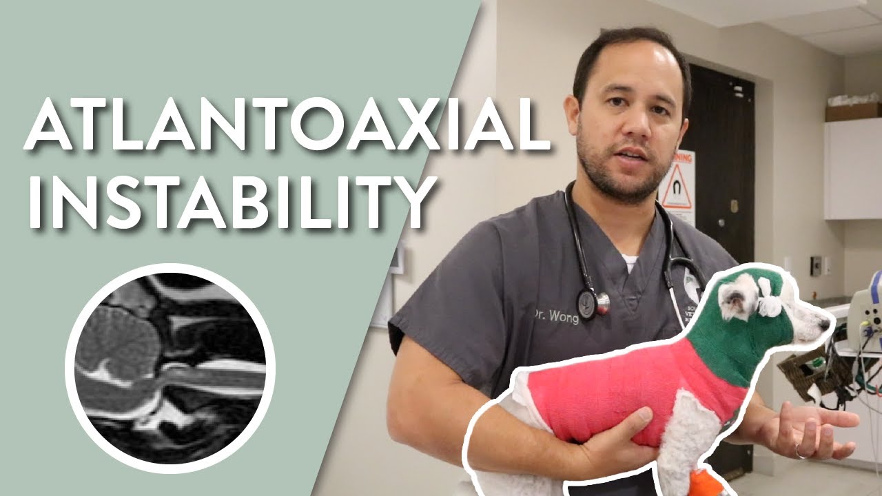
Image taken from the YouTube channel Southeast Veterinary Neurology , from the video titled What is Atlantoaxial Instability in Dogs? || Southeast Veterinary Neurology .
Atlantoaxial Luxation (AAL), a term that can sound daunting to any dog owner, refers to an instability in the upper cervical spine of our canine companions. Specifically, it involves the joint between the first two vertebrae, the atlas (C1) and the axis (C2). When this joint becomes unstable, it can lead to a range of neurological problems, significantly impacting a dog’s quality of life.
This guide aims to provide dog owners with a comprehensive understanding of AAL, covering its causes, diagnosis, treatment options, and long-term management strategies. Empowering you with knowledge is the first step in advocating for your dog’s health.
Defining Atlantoaxial Luxation
Atlantoaxial Luxation occurs when there is abnormal movement or displacement between the atlas (C1) and axis (C2) vertebrae. These vertebrae are critical for head and neck movement.
This instability can put pressure on the spinal cord, which runs through the vertebral column, leading to neurological deficits. The severity of these deficits depends on the degree of compression and the duration of the condition.
The Vital Role of the Atlas (C1) and Axis (C2)
The atlas (C1) and axis (C2) are uniquely shaped vertebrae that allow for a wide range of head and neck motion. The atlas, lacking a vertebral body, articulates with the skull, allowing nodding movements. The axis, with its prominent dens (odontoid process), fits into the atlas, enabling rotational movements.
Ligaments normally hold these vertebrae securely together, preventing excessive movement. In AAL, these ligaments are either absent, weakened, or damaged, leading to instability.
The Importance of Early Detection and Treatment
Early detection and prompt treatment are crucial in managing AAL. The longer the spinal cord is compressed, the more severe and potentially irreversible the neurological damage can become.
Recognizing the early signs of AAL, such as neck pain or reluctance to move the head, is essential for seeking timely veterinary care.
Empowering Owners Through Knowledge
This guide is designed to equip dog owners with the knowledge necessary to understand AAL and actively participate in their dog’s care. From understanding the causes and recognizing the symptoms to navigating diagnostic procedures and treatment options, this guide aims to empower you to advocate for your dog’s health and well-being.
The ultimate goal is to enable informed decision-making in partnership with your veterinarian, ensuring the best possible outcome for your beloved canine companion.
The unique anatomy of the atlas and axis vertebrae allows our canine friends to explore the world with impressive neck flexibility. However, this intricate structure is also susceptible to instability, leading to Atlantoaxial Luxation (AAL). Understanding the root causes of this condition is crucial for prevention, early detection, and informed decision-making regarding your dog’s health.
What Causes Atlantoaxial Luxation in Dogs?
Atlantoaxial Luxation isn’t a single disease with one simple trigger. Instead, it arises from a complex interplay of factors, broadly categorized as either congenital (present at birth) or acquired (developing after birth, usually due to trauma).
Congenital vs. Acquired AAL: Two Distinct Pathways
The distinction between congenital and acquired AAL is paramount because it influences both the likelihood of the condition and the potential preventative measures.
Congenital AAL: A Developmental Imperfection
Congenital AAL stems from developmental abnormalities that occur as a puppy grows in the womb.
This means the ligaments and bony structures meant to stabilize the atlas and axis vertebrae don’t form correctly.
In some cases, the dens, the crucial projection on the axis that fits into the atlas, may be malformed or entirely absent.
This congenital absence or malformation of the dens results in an unstable joint from the start, predisposing the puppy to luxation even with minimal stress or trauma.
Acquired AAL: The Result of Trauma
Acquired AAL, on the other hand, develops due to trauma to the neck region.
This trauma can range from seemingly minor incidents like a fall from furniture to more severe events like being hit by a car.
The force of the impact can damage or rupture the ligaments holding the atlas and axis together, leading to instability.
While any dog can experience acquired AAL, those with pre-existing, subtle congenital weaknesses may be more vulnerable.
Breed Predisposition: A Genetic Component
While trauma can affect any dog, certain breeds have a significantly higher risk of developing AAL, pointing to a strong genetic predisposition.
Small Breeds, Higher Risk
Small breed dogs, particularly toy breeds, are disproportionately affected by AAL.
This is believed to be due to a combination of factors, including their delicate bone structures and a higher prevalence of certain genetic mutations that affect vertebral development.
High-Risk Breeds
Several breeds are recognized as being at higher risk for AAL:
- Yorkshire Terriers: This breed is commonly affected due to congenital abnormalities in the formation of the atlantoaxial joint.
- Chihuahuas: Similar to Yorkshire Terriers, Chihuahuas often present with congenital AAL.
- Pomeranians: Their small size and genetic predisposition make them more susceptible.
- Toy Poodles: Another small breed where congenital AAL is frequently diagnosed.
- Maltese: The breed has genetic lines predisposed to the condition.
While these breeds are at higher risk, it’s important to remember that AAL can occur in dogs of any breed or size.
Understanding the breed-specific risks allows owners and breeders to be more vigilant, seeking early diagnosis and implementing preventative measures when possible.
Acquired AAL might stem from a singular traumatic event, while congenital forms often manifest gradually. Regardless of the origin, recognizing the telltale signs of Atlantoaxial Luxation is paramount. The sooner you identify these symptoms, the faster you can seek veterinary intervention and potentially mitigate long-term neurological damage.
Recognizing the Signs: Symptoms of Atlantoaxial Luxation
Atlantoaxial Luxation (AAL) presents a spectrum of symptoms, reflecting the degree of spinal cord compression and the stability (or instability) of the atlantoaxial joint. These signs can range from subtle indicators of discomfort to severe neurological deficits, significantly impacting a dog’s mobility and overall well-being. Early recognition of these symptoms is crucial, as AAL is often a progressive condition. Prompt veterinary attention can make a significant difference in managing the condition and improving the dog’s prognosis.
Common Symptoms of AAL
The clinical presentation of AAL is diverse, but several common symptoms should raise suspicion. It’s important to remember that not all dogs will exhibit every symptom, and the severity can vary greatly.
-
Neck pain and stiffness: This is often one of the first and most noticeable signs. Affected dogs may cry out when their neck is touched or resist any attempts to manipulate their head and neck.
-
Reluctance to move the head: Dogs may avoid turning their head from side to side or up and down. They might turn their entire body instead of just their head to look at something.
-
Uncoordinated gait (ataxia): Ataxia, or loss of coordination, is a significant sign of neurological involvement. Dogs may appear wobbly, stagger, or have difficulty placing their feet correctly.
-
Weakness in the limbs: Limb weakness can manifest as an inability to support their weight, dragging of the paws, or difficulty rising from a lying position. This weakness can affect all four limbs or be more pronounced in the hind limbs.
-
Paralysis (in severe cases): Paralysis, the complete loss of movement, represents the most severe manifestation of AAL. It typically indicates significant spinal cord compression and requires immediate veterinary intervention.
The Progressive Nature of AAL
One of the critical aspects of AAL is its potential to worsen over time. Initially, symptoms might be mild and intermittent, leading owners to dismiss them as a minor strain or temporary discomfort.
However, as the atlantoaxial joint becomes increasingly unstable, the spinal cord endures more compression, leading to a gradual worsening of the symptoms. This progression can be rapid in some cases, particularly after a traumatic event.
It is crucial to recognize that even seemingly minor symptoms could indicate the early stages of AAL. Ignoring these early warning signs can lead to irreversible spinal cord damage and a poorer prognosis.
The Urgency of Veterinary Care
Given the progressive nature of AAL and the potential for severe neurological damage, seeking veterinary care at the first sign of trouble is paramount. A veterinarian can perform a thorough neurological examination, utilize diagnostic imaging (such as X-rays, CT scans, or MRI), and determine the underlying cause of the symptoms.
Early diagnosis and treatment are essential to stabilize the atlantoaxial joint, relieve spinal cord compression, and improve the dog’s chances of a successful recovery. Delaying veterinary care can lead to permanent disability and a diminished quality of life for your beloved companion.
Recognizing the potential symptoms of Atlantoaxial Luxation is the first step, but confirming the diagnosis requires a thorough and systematic approach. A definitive diagnosis isn’t solely based on observed symptoms; it involves a combination of careful clinical assessment and advanced imaging techniques, all orchestrated by a skilled veterinarian.
Diagnosis: Confirming Atlantoaxial Luxation
The diagnostic process for Atlantoaxial Luxation (AAL) is multifaceted, requiring a veterinarian’s expertise to interpret clinical signs and radiographic findings. This process aims to not only confirm the presence of AAL but also to assess the severity of the spinal cord compression, which is crucial for determining the most appropriate treatment plan. Let’s take a look at the steps involved.
The Veterinarian’s Crucial Role
The veterinarian acts as the central figure in diagnosing AAL. Their role extends beyond simply identifying the condition; they are responsible for:
-
Gathering a comprehensive history: This includes information about the dog’s breed, age, onset of symptoms, and any history of trauma.
-
Performing a thorough physical examination: Assessing the dog’s overall health and identifying any other potential contributing factors.
-
Interpreting diagnostic results: Synthesizing information from neurological examinations and imaging studies to reach an accurate diagnosis.
-
Developing a tailored treatment plan: Based on the diagnosis, severity of the condition, and the dog’s individual needs.
Diagnostic Procedures: A Step-by-Step Approach
Several diagnostic procedures are available to confirm AAL and assess its severity. These range from non-invasive neurological exams to advanced imaging techniques.
Neurological Examination: Assessing Reflexes and Motor Function
A neurological examination is a critical first step in the diagnostic process. It helps the veterinarian evaluate the function of the dog’s nervous system and pinpoint the location of any neurological deficits.
During the examination, the veterinarian will assess:
- Reflexes: Evaluating the dog’s reflexes helps determine the integrity of the spinal cord and peripheral nerves.
- Motor function: Observing the dog’s gait, posture, and ability to move its limbs to identify any weakness, incoordination, or paralysis.
- Pain perception: Assessing the dog’s response to stimuli to determine the extent of spinal cord compression.
- Cranial nerve function: Checking the nerves of the head to look for abnormalities.
Specific neurological deficits, such as decreased reflexes in the hind limbs or an abnormal gait, can strongly suggest spinal cord compression in the region of the atlantoaxial joint.
X-Rays: Initial Assessment of Vertebral Alignment
Radiographs, or X-rays, are often the initial imaging modality used to evaluate dogs suspected of having AAL. X-rays provide a basic assessment of the alignment of the atlas (C1) and axis (C2) vertebrae.
While X-rays can reveal a misalignment or instability of the atlantoaxial joint, they have limitations. They don’t visualize the spinal cord itself or other soft tissues surrounding the vertebrae. Therefore, further imaging studies are often necessary to confirm the diagnosis and assess the severity of spinal cord compression.
CT Scans: Providing Detailed Bone Structure Information
Computed Tomography (CT) scans offer a more detailed view of the bony structures of the atlantoaxial joint than X-rays. CT scans use X-rays to create cross-sectional images of the spine, allowing veterinarians to assess the:
- Degree of luxation: Precisely measuring the displacement of the atlas and axis vertebrae.
- Presence of fractures: Identifying any fractures or bone abnormalities that may be contributing to the instability.
- Extent of bony compression: Assessing how the luxation is impacting the space available for the spinal cord.
CT scans are particularly useful for surgical planning. The detailed images help surgeons visualize the anatomy of the atlantoaxial joint and plan the most effective surgical approach.
MRI: Visualizing the Spinal Cord and Soft Tissues
Magnetic Resonance Imaging (MRI) is considered the gold standard for diagnosing AAL because it provides the most detailed visualization of the spinal cord and surrounding soft tissues.
MRI uses magnetic fields and radio waves to create images of the spine, allowing veterinarians to assess:
- Spinal cord compression: Directly visualizing the extent to which the spinal cord is being compressed by the luxated vertebrae.
- Spinal cord damage: Identifying any signs of inflammation, swelling, or bleeding within the spinal cord.
- Ligament damage: Assessing the integrity of the ligaments that support the atlantoaxial joint.
- Disc disease: Ruling out differential diagnosis such as bulging or herniated intervertebral discs.
MRI is crucial for determining the prognosis and guiding treatment decisions. The information obtained from an MRI can help veterinarians determine whether surgery is necessary and what type of surgical procedure would be most appropriate.
Treatment Options: Surgery vs. Medical Management
Once a definitive diagnosis of Atlantoaxial Luxation (AAL) has been established, the critical decision of how to proceed with treatment arises. The therapeutic landscape for AAL encompasses two primary approaches: surgical intervention and medical management. Each pathway presents its own set of advantages, disadvantages, and considerations, demanding a careful evaluation to determine the optimal course of action for each individual canine patient. The decision isn’t always straightforward, hinging on factors like the severity of the luxation, the dog’s overall health status, and the financial realities of the owner.
Surgical Interventions: A Path to Stabilization
Surgical intervention is often considered the gold standard for treating AAL, particularly in severe cases where significant spinal cord compression is present. The overarching goal of surgery is to stabilize the atlantoaxial joint, thereby preventing further injury to the spinal cord and alleviating neurological deficits. Several surgical techniques are employed, each tailored to address the specific anatomical abnormalities present.
Fusion: Solidifying the Instability
Atlantoaxial fusion aims to create a permanent, stable connection between the atlas (C1) and axis (C2) vertebrae. This involves using bone grafts and surgical implants, such as screws and plates, to immobilize the joint. Over time, the bone graft integrates with the vertebrae, resulting in a solid, fused segment.
This procedure effectively eliminates the abnormal movement that causes spinal cord compression. Fusion is often recommended for dogs with severe instability or those who have not responded to medical management.
Dorsal Laminectomy: Decompressing the Spinal Cord
Dorsal laminectomy is a surgical procedure designed to relieve pressure on the spinal cord. It involves removing a portion of the vertebral lamina (the bony arch of the vertebra) to create more space for the spinal cord. This can be particularly beneficial in cases where bone fragments or other tissue are impinging on the spinal cord.
Laminectomy is often performed in conjunction with fusion to achieve both stabilization and decompression.
Shunts and Implants: Advanced Solutions
In some instances, particularly those involving complex anatomical abnormalities, specialized shunts or other implants may be utilized. These devices can provide additional support to the atlantoaxial joint or help to redirect pressure away from the spinal cord. The specific type of implant used will depend on the individual dog’s condition and the surgeon’s expertise.
Medical Management: A Conservative Approach
Medical management represents a non-surgical approach to treating AAL. It focuses on alleviating pain, preventing further injury, and allowing the body to heal naturally. Medical management is typically reserved for mild cases of AAL or for dogs who are not suitable candidates for surgery due to underlying health conditions or financial constraints.
Pain Management: Comfort and Relief
Pain management is a cornerstone of medical management for AAL. Medications such as non-steroidal anti-inflammatory drugs (NSAIDs) and opioids are commonly used to reduce pain and inflammation. The specific medication and dosage will be determined by the veterinarian based on the dog’s individual needs and response to treatment.
Strict Cage Rest: Limiting Movement
Strict cage rest is essential to prevent further injury to the unstable atlantoaxial joint. The dog should be confined to a small, comfortable space, such as a crate or pen, to limit movement and minimize the risk of exacerbating the luxation.
Neck Brace Support
A neck brace can provide external support to the neck and help to stabilize the atlantoaxial joint. This can reduce pain and prevent further injury, particularly during the initial stages of treatment.
The brace must be properly fitted by a veterinarian or veterinary technician to ensure optimal support and comfort.
Factors Influencing Treatment Choice: A Holistic Evaluation
The decision between surgical intervention and medical management is complex and multifaceted. Several key factors must be considered to determine the most appropriate course of action for each individual dog.
-
Severity of the Condition: Dogs with severe spinal cord compression or instability are generally better candidates for surgery.
-
Overall Health: Dogs with significant underlying health conditions may not be able to tolerate the risks associated with surgery.
-
Cost: Surgical intervention is typically more expensive than medical management, which can be a significant factor for many owners.
Ultimately, the treatment decision should be made in consultation with a qualified veterinarian who can carefully evaluate all relevant factors and provide personalized recommendations. A frank discussion about the potential benefits, risks, and costs of each treatment option is essential to ensure that the owner is fully informed and able to make the best decision for their beloved companion.
Surgery or medical management represents only the initial step in addressing Atlantoaxial Luxation. The journey to recovery extends far beyond the operating room or the administration of medication, demanding a dedicated and comprehensive approach to post-treatment care and rehabilitation. This phase is critical in maximizing the chances of a successful outcome and restoring a fulfilling quality of life for affected dogs.
Post-Treatment Care and Rehabilitation
The period following either surgical intervention or medical management for AAL is crucial. It sets the stage for long-term recovery and well-being. A well-structured post-treatment plan addresses pain, restores physical function, and prevents potential complications. Neglecting this phase can significantly impede progress and diminish the overall success of the chosen treatment strategy.
Pain Management After AAL Treatment
Effective pain management is paramount in the immediate aftermath of surgery or the initiation of medical management. Dogs recovering from AAL often experience significant discomfort. Addressing this pain not only improves their comfort but also facilitates participation in rehabilitation exercises.
Veterinarians typically prescribe a combination of pain medications, including:
-
Opioids: For short-term, acute pain relief following surgery.
-
Non-Steroidal Anti-Inflammatory Drugs (NSAIDs): To manage inflammation and provide ongoing pain relief.
-
Adjunct Analgesics: Such as gabapentin or amantadine, which can help with neuropathic pain.
Regular monitoring of pain levels is essential. Owners should communicate any signs of discomfort to their veterinarian. Dosage adjustments or changes in medication may be necessary to ensure optimal pain control. Furthermore, non-pharmacological approaches, such as acupuncture or laser therapy, may be considered as adjuncts to traditional pain medications.
The Role of Rehabilitation in AAL Recovery
Rehabilitation plays a vital role in restoring strength, coordination, and mobility in dogs recovering from AAL. A tailored rehabilitation program, designed in consultation with a veterinary rehabilitation specialist, can significantly improve the outcome.
Physical Therapy for Strength and Coordination
Physical therapy exercises are fundamental to regaining lost function. These exercises aim to strengthen weakened muscles, improve balance, and restore a normal gait. Common physical therapy techniques include:
-
Passive Range of Motion (PROM) Exercises: Gently moving the limbs through their natural range of motion to prevent stiffness and improve joint flexibility.
-
Strengthening Exercises: Using resistance bands or other tools to build muscle strength in the limbs and core.
-
Balance and Coordination Exercises: Such as standing on a wobble board or walking over uneven surfaces, to improve proprioception (awareness of body position in space).
Hydrotherapy: A Low-Impact Option
Hydrotherapy, or underwater treadmill therapy, offers a low-impact exercise option that is particularly beneficial for dogs recovering from AAL. The buoyancy of water reduces stress on the joints, allowing for greater range of motion and reduced pain during exercise. Hydrotherapy can improve cardiovascular fitness, muscle strength, and overall mobility.
Ongoing Monitoring and Treatment Plan Adjustments
Recovery from AAL is not a linear process. It requires continuous monitoring and a willingness to adjust the treatment plan as needed. Regular veterinary check-ups are crucial to assess progress, identify any potential complications, and make necessary modifications to the pain management or rehabilitation protocols.
Owners should be vigilant in observing their dog’s behavior and reporting any changes to their veterinarian. Subtle signs of discomfort or decreased mobility may indicate the need for adjustments to the medication or therapy regimen. A collaborative approach between the owner, veterinarian, and rehabilitation specialist is essential to ensure the best possible outcome for the dog. Flexibility and responsiveness are key to navigating the complexities of AAL recovery.
Prognosis and Long-Term Management of Atlantoaxial Luxation in Dogs
The diagnosis of Atlantoaxial Luxation (AAL) in a beloved canine companion can be a daunting experience. While the initial treatment, whether surgical or medical, is crucial, understanding the long-term outlook and management strategies is equally vital for ensuring the best possible quality of life for your dog. The prognosis for dogs with AAL varies depending on several factors, but with diligent care and proactive management, many can live comfortable and fulfilling lives.
Factors Influencing Prognosis
The prognosis for a dog diagnosed with AAL is not a fixed entity. It is influenced by a constellation of factors that must be carefully considered. These factors help veterinarians and owners make informed decisions about ongoing care and expectations.
-
Severity of the Luxation: The degree of spinal cord compression plays a significant role. Dogs with mild to moderate compression often have a better prognosis than those with severe compression and paralysis.
-
Age and Overall Health: Younger dogs often demonstrate greater resilience and adaptability. The presence of other underlying health conditions can complicate recovery and impact the long-term prognosis.
-
Treatment Response: The effectiveness of the chosen treatment, whether surgical or medical, is a critical determinant. A positive response to initial therapy significantly improves the long-term outlook. Early intervention is crucial.
-
Presence of Neurological Deficits: Dogs that retain some motor function and sensation tend to have a more favorable prognosis compared to those with complete paralysis.
Long-Term Management Considerations
Effective long-term management is essential for maximizing the quality of life for dogs with AAL. This involves a multi-faceted approach focused on pain control, regular monitoring, and environmental modifications.
Ongoing Pain Management
Pain management is a cornerstone of long-term care. Although the initial pain associated with AAL may subside after treatment, chronic pain can persist in some cases.
-
Medication: Veterinarians may prescribe ongoing pain medication, such as NSAIDs, gabapentin, or amantadine, to manage chronic pain. Regular monitoring for potential side effects is essential.
-
Alternative Therapies: Acupuncture, laser therapy, and massage can be valuable adjuncts to traditional pain medication. These therapies can help reduce inflammation, alleviate muscle tension, and improve overall comfort.
Regular Veterinary Check-Ups
Consistent monitoring by a veterinarian is crucial. These check-ups allow for early detection of any potential problems and timely adjustments to the management plan.
-
Neurological Assessments: Regular neurological evaluations can help track the dog’s progress and identify any changes in neurological function.
-
Imaging Studies: Periodic X-rays or other imaging studies may be necessary to monitor the stability of the atlantoaxial joint and assess for any signs of recurrence or complications.
Environmental Modifications
Creating a safe and supportive environment is vital for preventing further injury and promoting well-being.
-
Assistive Devices: Depending on the dog’s mobility, assistive devices such as harnesses or wheelchairs can help improve their ability to move around.
-
Home Adaptations: Ramps or steps can make it easier for dogs to access furniture or navigate stairs. Non-slip flooring can also help prevent falls. Soft bedding can prevent pressure sores if mobility is reduced.
-
Controlled Exercise: While activity is important, it should be carefully controlled to avoid overexertion or potential injury. Short, frequent walks on leash are generally recommended.
Potential for Recurrence or Complications
While treatment can significantly improve the quality of life for dogs with AAL, the potential for recurrence or complications remains.
-
Instability: The atlantoaxial joint may become unstable again, leading to a recurrence of symptoms. This is more likely to occur in cases where surgical fusion was not performed or was unsuccessful.
-
Progressive Spinal Cord Damage: Despite treatment, some dogs may experience progressive spinal cord damage, leading to worsening neurological deficits.
-
Arthritis: Over time, arthritis can develop in the atlantoaxial joint, contributing to pain and stiffness.
-
Pressure Sores: Dogs with decreased mobility are at risk of developing pressure sores. Regular turning and soft bedding can help prevent these sores.
Owners must be vigilant in monitoring their dogs for any signs of recurrence or complications. Prompt veterinary attention is essential to address these issues and prevent further decline.
Effective long-term management is essential for maximizing the quality of life for dogs diagnosed with Atlantoaxial Luxation. However, navigating the complexities of AAL requires a strong partnership between you and your veterinarian. Choosing the right veterinary professional and fostering open communication are critical components of ensuring the best possible care for your canine companion.
Working with Your Veterinarian: Seeking the Best Care
Finding a veterinarian who is not only skilled but also experienced in managing Atlantoaxial Luxation (AAL) is paramount. This condition demands a nuanced understanding of neurological disorders, advanced imaging techniques, and both surgical and medical management options.
The Importance of Expertise
Not all veterinarians have extensive experience with AAL.
Look for a veterinary neurologist or a surgeon with a strong track record in spinal surgeries.
Experience matters. An experienced veterinarian will be better equipped to:
- Accurately diagnose AAL.
- Develop a tailored treatment plan.
- Anticipate and manage potential complications.
Finding the Right Veterinary Professional
Identifying a qualified veterinarian requires proactive effort. Start by:
- Asking your current veterinarian for a referral to a specialist.
- Contacting veterinary teaching hospitals in your region.
- Searching online directories of board-certified veterinary neurologists and surgeons.
Consider leveraging resources like the American Animal Hospital Association (AAHA).
While AAHA does not directly certify individual veterinarians in specialties, they do accredit veterinary hospitals that adhere to high standards of care.
These hospitals often have teams of experienced professionals, including specialists who can provide expert care for dogs with AAL.
AAHA and Veterinary Guidelines
AAHA plays a crucial role in setting standards for veterinary practice.
Their guidelines cover a wide range of topics, including:
- Pain management
- Surgical protocols
- Rehabilitation strategies
While not specifically focused on AAL, these guidelines ensure that accredited hospitals provide evidence-based, high-quality care.
Veterinarians who adhere to AAHA standards are committed to staying up-to-date with the latest advancements in veterinary medicine.
This commitment translates to better outcomes for your dog.
Open Communication: A Cornerstone of Care
Once you’ve found a veterinarian you trust, fostering open and honest communication is key.
Be prepared to ask questions, express concerns, and actively participate in your dog’s care.
Effective communication ensures that:
- You fully understand the diagnosis, treatment options, and prognosis.
- Your veterinarian is aware of your dog’s specific needs and your personal preferences.
- You can work together to make informed decisions about your dog’s care.
Don’t hesitate to:
- Ask for clarification on any medical terms or procedures you don’t understand.
- Share your observations about your dog’s behavior and response to treatment.
- Discuss your concerns about the cost of treatment and explore available financial assistance options.
By actively engaging in the communication process, you can build a strong partnership with your veterinarian and advocate for the best possible care for your beloved dog.
FAQs: Atlantoaxial Luxation in Dogs
Here are some frequently asked questions about atlantoaxial luxation in dogs, designed to help you better understand this condition.
What exactly is atlantoaxial luxation in dogs?
Atlantoaxial luxation is a spinal condition where the first two vertebrae in the neck (atlas and axis) become unstable. This instability can cause spinal cord compression, leading to neurological problems. Atlantoaxial luxation dog cases often stem from congenital abnormalities or trauma.
How is atlantoaxial luxation diagnosed?
Diagnosis typically involves a neurological exam, X-rays, and sometimes advanced imaging like CT scans or MRI. Vets look for misalignment of the vertebrae and signs of spinal cord compression, which are key indicators of atlantoaxial luxation dog.
What are the treatment options for atlantoaxial luxation?
Treatment options range from conservative management (rest, pain medication, bracing) to surgical stabilization. Surgery aims to fuse the atlas and axis, preventing further movement and relieving pressure on the spinal cord. The best approach for an atlantoaxial luxation dog depends on the severity and individual circumstances.
What is the long-term outlook after treatment?
The prognosis depends on the severity of the luxation and the chosen treatment method. With successful surgery and rehabilitation, many dogs with atlantoaxial luxation dog can regain function and live comfortable lives. However, some dogs may experience residual neurological deficits.
Navigating atlantoaxial luxation dog can feel overwhelming, but remember you’re not alone! Armed with this knowledge, you’re in a better position to advocate for your furry friend’s health and happiness.
